Explore
 Validate
 Learn
Learn Western blot
Antibody data
- Antibody Data
- Antigen structure
- References [5]
- Comments [0]
- Validations
- Western blot [2]
- Immunohistochemistry [1]
- Other assay [2]
Submit
Validation data
Reference
Comment
Report error
- Product number
- PA5-19479 - Provider product page

- Provider
- Invitrogen Antibodies
- Product name
- E-cadherin Polyclonal Antibody
- Antibody type
- Polyclonal
- Antigen
- Synthetic peptide
- Description
- A diffuse signal may be seen at higher concentrations (5-10 µg/mL) in ICC/IF. For Western Blot, this antibody has non-specific bands at 40 kDa,65 kDa,75 kDa and 90 kDa. This antibody is predicted to react with chicken, cow and Xenopus laevis based on sequence homology. This antibody may not perform well in ICC/IF using mouse cells.
- Reactivity
- Human, Mouse, Rat
- Host
- Rabbit
- Isotype
- IgG
- Vial size
- 100 µg
- Concentration
- 1 mg/mL
- Storage
- -20°C or -80°C if preferred
Submitted references Deciphering the effects of PYCR1 on cell function and its associated mechanism in hepatocellular carcinoma.
IL-20 Cytokines Are Involved in Epithelial Lesions Associated with Virus-Induced COPD Exacerbation in Mice.
Differential expression of cell-cell junction proteins in the testis, epididymis, and ductus deferens of domestic turkeys (Meleagris gallopavo) with white and yellow semen.
Functional Subclone Profiling for Prediction of Treatment-Induced Intratumor Population Shifts and Discovery of Rational Drug Combinations in Human Glioblastoma.
Analysis of in vivo single cell behavior by high throughput, human-in-the-loop segmentation of three-dimensional images.
Xu Y, Zuo W, Wang X, Zhang Q, Gan X, Tan N, Jia W, Liu J, Li Z, Zhou B, Zhao D, Xie Z, Tan Y, Zheng S, Liu C, Li H, Chen Z, Yang X, Huang Z
International journal of biological sciences 2021;17(9):2223-2239
International journal of biological sciences 2021;17(9):2223-2239
IL-20 Cytokines Are Involved in Epithelial Lesions Associated with Virus-Induced COPD Exacerbation in Mice.
Le Roux M, Ollivier A, Kervoaze G, Beke T, Gillet L, Pichavant M, Gosset P
Biomedicines 2021 Dec 5;9(12)
Biomedicines 2021 Dec 5;9(12)
Differential expression of cell-cell junction proteins in the testis, epididymis, and ductus deferens of domestic turkeys (Meleagris gallopavo) with white and yellow semen.
Pardyak L, Kaminska A, Brzoskwinia M, Hejmej A, Kotula-Balak M, Jankowski J, Ciereszko A, Bilinska B
Poultry science 2020 Jan;99(1):555-566
Poultry science 2020 Jan;99(1):555-566
Functional Subclone Profiling for Prediction of Treatment-Induced Intratumor Population Shifts and Discovery of Rational Drug Combinations in Human Glioblastoma.
Reinartz R, Wang S, Kebir S, Silver DJ, Wieland A, Zheng T, Küpper M, Rauschenbach L, Fimmers R, Shepherd TM, Trageser D, Till A, Schäfer N, Glas M, Hillmer AM, Cichon S, Smith AA, Pietsch T, Liu Y, Reynolds BA, Yachnis A, Pincus DW, Simon M, Brüstle O, Steindler DA, Scheffler B
Clinical cancer research : an official journal of the American Association for Cancer Research 2017 Jan 15;23(2):562-574
Clinical cancer research : an official journal of the American Association for Cancer Research 2017 Jan 15;23(2):562-574
Analysis of in vivo single cell behavior by high throughput, human-in-the-loop segmentation of three-dimensional images.
Chiang M, Hallman S, Cinquin A, de Mochel NR, Paz A, Kawauchi S, Calof AL, Cho KW, Fowlkes CC, Cinquin O
BMC bioinformatics 2015 Nov 25;16:397
BMC bioinformatics 2015 Nov 25;16:397
No comments: Submit comment
Supportive validation
- Submitted by
- Invitrogen Antibodies (provider)
- Main image

- Experimental details
- Western blot analysis of Mouse Heart Tissue Lysate using Product # PA5-19479, pan Cadherin primary antibody at a dilution of 1 µg/mL (lane 1). Staining of Human Heart Tissue Lysate at a dilution of 1 µg/mL (lane 2). Blot treated with a secondary HRP-conjugated Goat polyclonal anti-Rabbit antibody was used at a dilution of 1:3000.

Supportive validation
- Submitted by
- Invitrogen Antibodies (provider)
- Main image

- Experimental details
- Immunohistochemical (formalin-fixed, paraffin-embedded) staining of Human Hippocampus tissue using Product # PA5-19479, anti-Pan Cadherin antibody. Primary antibody was used at a concentration of 1 µg/mL and exposed for 8 mins at room temp. The sample was pretreated using heat mediated antigen retrieval with Sodium Citrate Buffer (pH6/20mins). The detection method was a HRP conjugated polymer, DAB chromogen and the sample was counterstained with haematoxylin and mounted with DPX.

Supportive validation
- Submitted by
- Invitrogen Antibodies (provider)
- Main image

- Experimental details
- Occludin, ZO-1, Cx43, N- and E-cadherin, and beta-catenin mRNA (A to E) and protein (A' to E') expression in yellow semen syndrome (YSS) and white normal semen (WNS) testes, epididymides, and ductuli deferentes. Representative quantitative real-time PCR analysis of occludin (A), ZO-1 (B), Cx43 (C), N- and E-cadherin (D), and beta-catenin (E) mRNA expression in YSS and WNS testes, epididymides, and ductuli deferentes. As an internal control, beta-actin mRNA level was measured. Relative quantification (RQ) is expressed as mean +- standard deviation (SD). Data were obtained from 3 separate experiments. Asterisks indicate statistically significant differences (* P < 0.05, ** P < 0.01, *** P < 0.001; Mann-Whitney U-test). YSS turkeys (n = 6) and WNS turkeys (n = 6). Representative western blots and relative expression of occludin (A'), ZO-1 (B'), Cx43 (C'), N- and E-cadherin (D'), and beta-catenin (E') proteins in YSS and WNS testes, epididymides, and ductuli deferentes. Densitometric analysis of protein content was normalized against the corresponding beta-actin. Protein levels within the WNS tissues were arbitrary set as 1. Data obtained from 3 separate analyses are expressed as mean +- SD. Asterisks indicate statistically significant differences (* P < 0.05, ** P < 0.01, *** P < 0.001; Mann-Whitney U-test). N = 5 each for YSS and WNS tissues.

- Submitted by
- Invitrogen Antibodies (provider)
- Main image

- Experimental details
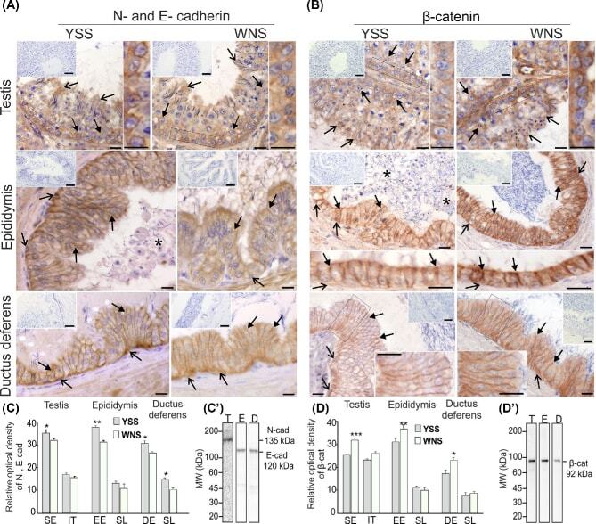
- Figure 3 Qualitative (A and B) and quantitative (C and D) analysis of immunohistochemical staining for N- and E-cadherin (A) and beta-catenin (B). Representative micrographs of yellow semen syndrome (YSS) and white normal semen (WNS) testes, epididymides, and ductuli deferentes. Counterstaining was performed with Mayer's haematoxylin. Scale bars are 10 mum. Frames indicate the location of the higher magnification view. (A and B) Similar staining pattern for N- and E-cadherin (A) and beta-catenin (B) were observed in the basal compartment at the blood-testis barrier (BTB) (arrows) and in the adluminal compartment of seminiferous epithelium (open arrows) as well as in the lateral plasma membranes of adjacent epididymal principal cells (arrows) and between principal and basal cells (open arrows), and in the lateral plasma membranes of adjacent ductal columnar cells (arrows) and between columnar and basal cells (open arrows) of YSS and WNS epididymides and ductuli deferentes. Note increased N- and E-cadherin and reduced beta-catenin staining intensity at the BTB of YSS testis (see higher magnification views in A and B) and of YSS epididymis and ductus deferens sections (see higher magnification views in B). Note weaker staining for beta-catenin between epithelial principal cells (arrows) and principal and basal cells (open arrows) along the entire YSS epididymis length (see higher magnification inserts in B). Note also sloughed germ cells admixed with mature spermatozoa in YSS ep