Explore
 Validate
 Learn
Learn Western blot
 ELISA
ELISAAntibody data
- Antibody Data
- Antigen structure
- References [2]
- Comments [0]
- Validations
- Western blot [2]
- Immunohistochemistry [1]
- Flow cytometry [1]
- Other assay [3]
Submit
Validation data
Reference
Comment
Report error
- Product number
- MA5-15711 - Provider product page

- Provider
- Invitrogen Antibodies
- Product name
- E-cadherin Monoclonal Antibody (7H12)
- Antibody type
- Monoclonal
- Antigen
- Purifed from natural sources
- Description
- MA5-15711 targets CDH1 in indirect ELISA, FACS, IHC, and WB applications and shows reactivity with Human, mouse, and Non-human primate samples. The MA5-15711 immunogen is purified recombinant fragment of human CDH1 expressed in E. Coli. MA5-15711 detects CDH1 which has a predicted molecular weight of approximately 135kDa.
- Reactivity
- Human, Mouse
- Host
- Mouse
- Isotype
- IgG
- Antibody clone number
- 7H12
- Vial size
- 100 µL
- Concentration
- Conc. Not Determined
- Storage
- Store at 4°C short term. For long term storage, store at -20°C, avoiding freeze/thaw cycles.
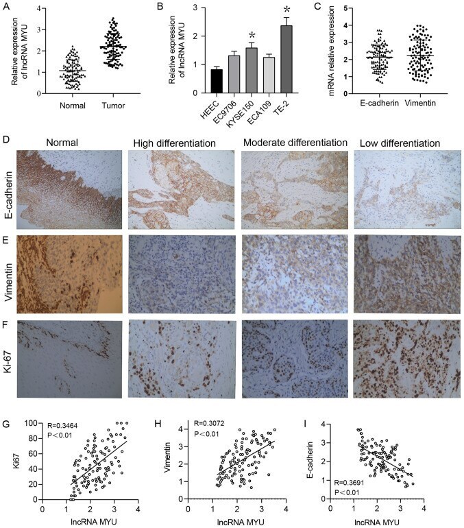
Submitted references Upregulation of long non-coding RNA MYU promotes proliferation, migration and invasion of esophageal squamous cell carcinoma cells.
Ablation of Doublecortin-Like Kinase 1 in the Colonic Epithelium Exacerbates Dextran Sulfate Sodium-Induced Colitis.
Gu S, Qian L, Liu Y, Miao J, Shen H, Zhang S, Mao G
Experimental and therapeutic medicine 2021 Jun;21(6):644
Experimental and therapeutic medicine 2021 Jun;21(6):644
Ablation of Doublecortin-Like Kinase 1 in the Colonic Epithelium Exacerbates Dextran Sulfate Sodium-Induced Colitis.
Qu D, Weygant N, May R, Chandrakesan P, Madhoun M, Ali N, Sureban SM, An G, Schlosser MJ, Houchen CW
PloS one 2015;10(8):e0134212
PloS one 2015;10(8):e0134212
No comments: Submit comment
Supportive validation
- Submitted by
- Invitrogen Antibodies (provider)
- Main image

- Experimental details
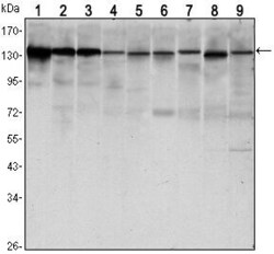
- Western blot analysis of CD324/E Cadherin using CD324/E Cadherin monoclonal antibody (Product # MA5-15711) in LNCAP (1) A431 (2), DU145 (3), PC-3 (4), MCF-7 (5), PC-12 (6), NIH/3T3 (7), C6 (8) and COS-7 (9) cell lysate.

- Submitted by
- Invitrogen Antibodies (provider)
- Main image
- Experimental details
- Western blot analysis of CD324/E Cadherin using CD324/E Cadherin monoclonal antibody (Product # MA5-15711) in LNCAP (1) A431 (2), DU145 (3), PC-3 (4), MCF-7 (5), PC-12 (6), NIH/3T3 (7), C6 (8) and COS-7 (9) cell lysate.

Supportive validation
- Submitted by
- Invitrogen Antibodies (provider)
- Main image
- Experimental details
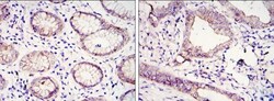
- Immunohistochemical analysis of paraffin-embedded gastric cancer tissues (left) and lung cancer tissues (right) using CD324/E Cadherin monoclonal antibody (Product # MA5-15711) followed with DAB staining.

Supportive validation
- Submitted by
- Invitrogen Antibodies (provider)
- Main image

- Experimental details
- Flow cytometric analysis of HeLa cells using CD324/E Cadherin monoclonal antibody (Product # MA5-15711) (green) and negative control (purple).

Supportive validation
- Submitted by
- Invitrogen Antibodies (provider)
- Main image

- Experimental details
- Fig 1 Characterization of intestinal epithelial deletion of Dclk1 under normal conditions. A. There is no detectable Dclk1 in the colonic epithelia of Villin Cre ;Dclk1 f/f mice. Colonic tissues stained with anti-Dclk1 antibody (Brown) under normal conditions (200x magnification). B . Dclk1 mRNA levels were dramatically decreased in the Villin Cre ;Dclk1 f/f mice; C . The expression levels of Dclk1 and TJPs in Dclk1 f/f and Villin Cre ;Dclk1 f/f mice; D . Intestinal mucosal integrity was impaired in the Villin Cre ;Dclk1 f/f mice. FITC-Dextran level in the serum of each mouse was determined 4 h after gavage (n = 3 for each group, *p

- Submitted by
- Invitrogen Antibodies (provider)
- Main image

- Experimental details
- Fig 2 Deletion of Dclk1 exacerbates colonic barrier dysfunction following DSS treatment. A . Experimental plan; B . H&E staining demonstrated normal histology in Villin Cre ;Dclk1 f/f and Dclk1 f/f mice at baseline and confirmed a significant inflammatory response following DSS treatment (200x magnification) C . Western blot analysis of tight junction and adherens junction proteins demonstrated a decrease in Claudin-1, Claudin-7, and E-cadherin in the Villin Cre ;Dclk1 f/f mice after DSS treatment; D. FITC-Dextran levels in the serum of each mouse were determined 4 h after gavage in baseline and post DSS treatment (n = 5 for post DSS groups, *p

- Submitted by
- Invitrogen Antibodies (provider)
- Main image
- Experimental details
- Figure 1 Expression of lncRNA MYU, E-cadherin, Vimentin and Ki-67 in ESCC tissues. (A) The relative expression of lncRNA MYU was increased in ESCC tissues compared with that in normal tissues. GAPDH was used as a loading control. (B) Reverse transcription-quantitative PCR analysis of lncRNA MYU expression in 3 ESCC cell lines and HEECs. The relative expression level of lncRNA MYU was significantly upregulated in TE-2 cells. * P