Explore
 Validate
 Learn
Learn Western blot
 Flow cytometry
Antibody data
- Antibody Data
- Antigen structure
- References [6]
- Comments [0]
- Validations
- Western blot [1]
- Immunocytochemistry [1]
- Immunohistochemistry [2]
Submit
Validation data
Reference
Comment
Report error
- Product number
- MAB18381 - Provider product page

- Provider
- R&D Systems
- Product name
- Human E-Cadherin Antibody
- Antibody type
- Monoclonal
- Description
- Protein A or G purified from hybridoma culture supernatant. Detects human E-Cadherin in direct ELISAs.
- Reactivity
- Human
- Host
- Mouse
- Conjugate
- Unconjugated
- Antigen sequence
P12830- Isotype
- IgG
- Antibody clone number
- 180224
- Vial size
- 100 ug
- Concentration
- LYOPH
- Storage
- Use a manual defrost freezer and avoid repeated freeze-thaw cycles. 12 months from date of receipt, -20 to -70 °C as supplied. 1 month, 2 to 8 °C under sterile conditions after reconstitution. 6 months, -20 to -70 °C under sterile conditions after reconstitution.
Submitted references Semaphorin 3A induces mesenchymal-stem-like properties in human periodontal ligament cells.
Generation of glucose-responsive, insulin-producing cells from human umbilical cord blood-derived mesenchymal stem cells.
Gram-positive bacteria enhance HIV-1 susceptibility in Langerhans cells, but not in dendritic cells, via Toll-like receptor activation.
Generation of pluripotent stem cells from adult human testis.
Expression of mesenchyme-specific gene HMGA2 in squamous cell carcinomas of the oral cavity.
Expression of mesenchyme-specific gene HMGA2 in squamous cell carcinomas of the oral cavity.
Wada N, Maeda H, Hasegawa D, Gronthos S, Bartold PM, Menicanin D, Fujii S, Yoshida S, Tomokiyo A, Monnouchi S, Akamine A
Stem cells and development 2014 Sep 15;23(18):2225-36
Stem cells and development 2014 Sep 15;23(18):2225-36
Generation of glucose-responsive, insulin-producing cells from human umbilical cord blood-derived mesenchymal stem cells.
Prabakar KR, Domínguez-Bendala J, Molano RD, Pileggi A, Villate S, Ricordi C, Inverardi L
Cell transplantation 2012;21(6):1321-39
Cell transplantation 2012;21(6):1321-39
Gram-positive bacteria enhance HIV-1 susceptibility in Langerhans cells, but not in dendritic cells, via Toll-like receptor activation.
Ogawa Y, Kawamura T, Kimura T, Ito M, Blauvelt A, Shimada S
Blood 2009 May 21;113(21):5157-66
Blood 2009 May 21;113(21):5157-66
Generation of pluripotent stem cells from adult human testis.
Conrad S, Renninger M, Hennenlotter J, Wiesner T, Just L, Bonin M, Aicher W, Bühring HJ, Mattheus U, Mack A, Wagner HJ, Minger S, Matzkies M, Reppel M, Hescheler J, Sievert KD, Stenzl A, Skutella T
Nature 2008 Nov 20;456(7220):344-9
Nature 2008 Nov 20;456(7220):344-9
Expression of mesenchyme-specific gene HMGA2 in squamous cell carcinomas of the oral cavity.
Miyazawa J, Mitoro A, Kawashiri S, Chada KK, Imai K
Cancer research 2004 Mar 15;64(6):2024-9
Cancer research 2004 Mar 15;64(6):2024-9
Expression of mesenchyme-specific gene HMGA2 in squamous cell carcinomas of the oral cavity.
Miyazawa J, Mitoro A, Kawashiri S, Chada KK, Imai K
Cancer research 2004 Mar 15;64(6):2024-9
Cancer research 2004 Mar 15;64(6):2024-9
No comments: Submit comment
Supportive validation
- Submitted by
- R&D Systems (provider)
- Main image
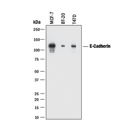
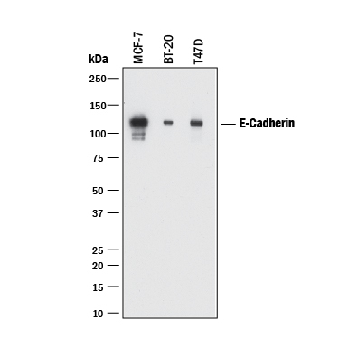
- Experimental details
- Detection of Human E-Cadherin by Western Blot. Western blot shows lysates of MCF-7 human breast cancer cell line, BT-20 human breast cancer cell line, and T47D human breast cancer cell line. PVDF membrane was probed with 2 µg/mL of Mouse Anti-Human E-Cadherin Monoclonal Antibody (Catalog # MAB18381) followed by HRP-conjugated Anti-Mouse IgG Secondary Antibody (Catalog # HAF018). A specific band was detected for E-Cadherin at approximately 120 kDa (as indicated). This experiment was conducted under reducing conditions and using Immunoblot Buffer Group 1.
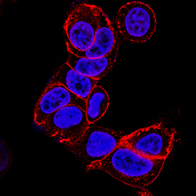
Supportive validation
- Submitted by
- R&D Systems (provider)
- Main image
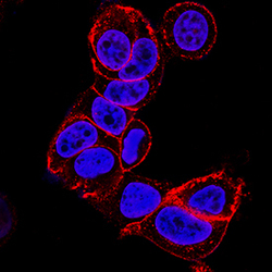
- Experimental details
- E-Cadherin in MCF-7 Human Cell Line. E-Cadherin was detected in immersion fixed MCF-7 human breast cancer cell line using Mouse Anti-Human E-Cadherin Monoclonal Antibody (Catalog # MAB18381) at 3 µg/mL for 3 hours at room temperature. Cells were stained using the NorthernLights™ 557-conjugated Anti-Mouse IgG Secondary Antibody (red; Catalog # NL007) and counterstained with DAPI (blue). Specific staining was localized to cell membrane. View our protocol for Fluorescent ICC Staining of Cells on Coverslips.
Supportive validation
- Submitted by
- R&D Systems (provider)
- Main image
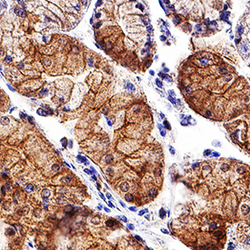
- Experimental details
- E-Cadherin in Human Stomach. E-Cadherin was detected in immersion fixed paraffin-embedded sections of human stomach using Mouse Anti-Human E-Cadherin Monoclonal Antibody (Catalog # MAB18381) at 5 µg/mL for 1 hour at room temperature followed by incubation with the Anti-Mouse IgG VisUCyte™ HRP Polymer Antibody (Catalog # VC001). Tissue was stained using DAB (brown) and counterstained with hematoxylin (blue). Specific staining was localized to plasma membrane. View our protocol for Chromogenic IHC Staining of Paraffin-embedded Tissue Sections.

- Submitted by
- R&D Systems (provider)
- Main image

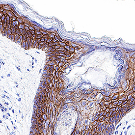
- Experimental details
- E-Cadherin in Human Skin. E-Cadherin was detected in immersion fixed paraffin-embedded sections of human skin using Mouse Anti-Human E-Cadherin Monoclonal Antibody (Catalog # MAB18381) at 0.5 µg/mL for 1 hour at room temperature followed by incubation with the Anti-Mouse IgG VisUCyte™ HRP Polymer Antibody (Catalog # VC001). Tissue was stained using DAB (brown) and counterstained with hematoxylin (blue). Specific staining was localized to plasma membrane. View our protocol for Chromogenic IHC Staining of Paraffin-embedded Tissue Sections.