Explore
 Validate
 Learn
Learn Immunohistochemistry
Antibody data
- Antibody Data
- Antigen structure
- References [2]
- Comments [0]
- Validations
- Immunohistochemistry [1]
Submit
Validation data
Reference
Comment
Report error
- Product number
- 600-401-116-0.1 - Provider product page

- Provider
- Rockland Immunochemicals, Inc.
- Proper citation
- Rockland Cat#600-401-116-0.1, RRID:AB_2134066
- Product name
- Anti-Laminin (RABBIT) Antibody - 600-401-116-0.1
- Antibody type
- Polyclonal
- Vial size
- 100 µl
Submitted references Perlecan and vascular endothelial growth factor-encoding DNA-loaded chitosan scaffolds promote angiogenesis and wound healing.
Oxidation modifies the structure and function of the extracellular matrix generated by human coronary artery endothelial cells.
Lord MS, Ellis AL, Farrugia BL, Whitelock JM, Grenett H, Li C, O'Grady RL, DeCarlo AA
Journal of controlled release : official journal of the Controlled Release Society 2017 Mar 28;250:48-61
Journal of controlled release : official journal of the Controlled Release Society 2017 Mar 28;250:48-61
Oxidation modifies the structure and function of the extracellular matrix generated by human coronary artery endothelial cells.
Chuang CY, Degendorfer G, Hammer A, Whitelock JM, Malle E, Davies MJ
The Biochemical journal 2014 Apr 15;459(2):313-22
The Biochemical journal 2014 Apr 15;459(2):313-22
No comments: Submit comment
Supportive validation
- Submitted by
- Rockland Immunochemicals, Inc. (provider)
- Main image
- Experimental details
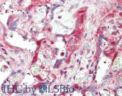
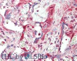
- Immunohistochemistry of rabbit anti-Laminin antibody. Tissue: skin. Fixation: formalin fixed paraffin embedded. Antigen retrieval: not required. Primary antibody: Anti-Laminin at 5 µg/mL for 1 h at RT. Secondary antibody: Peroxidase rabbit secondary antibody at 1:10,000 for 45 min at RT. Staining: Laminin as precipitated red signal with hematoxylin purple nuclear counterstain.
- Validation comment
- Immunohistochemistry