 Explore
 Validate
 Learn
Learn Western blot
 Immunohistochemistry
Antibody data
- Antibody Data
- Antigen structure
- References [6]
- Comments [0]
- Validations
- Immunohistochemistry [1]
Submit
Validation data
Reference
Comment
Report error
- Product number
- BAF459 - Provider product page

- Provider
- R&D Systems
- Product name
- Mouse Osteoprotegerin/TNFRSF11B Biotinylated Antibody
- Antibody type
- Polyclonal
- Description
- Antigen Affinity-purified. Detects mouse Osteoprotegerin/TNFRSF11B in ELISAs and Western blots. In sandwich immunoassays, less than 0.2% cross-reactivity with recombinant mouse RANK is observed.
- Reactivity
- Mouse
- Host
- Goat
- Conjugate
- Biotin
- Antigen sequence
Q6PI12- Isotype
- IgG
- Vial size
- 50 ug
- Concentration
- LYOPH
- Storage
- Use a manual defrost freezer and avoid repeated freeze-thaw cycles. 12 months from date of receipt, -20 to -70 °C as supplied. 1 month, 2 to 8 °C under sterile conditions after reconstitution. 6 months, -20 to -70 °C under sterile conditions after reconstitution.
Submitted references Osteoprotegerin-Mediated Homeostasis of Rank+ Thymic Epithelial Cells Does Not Limit Foxp3+ Regulatory T Cell Development.
OPG/RANKL/RANK axis is a critical inflammatory signaling system in ischemic brain in mice.
Cytokine-induced osteoprotegerin expression protects pancreatic beta cells through p38 mitogen-activated protein kinase signalling against cell death.
Therapeutic relevance of osteoprotegerin gene therapy in osteosarcoma: blockade of the vicious cycle between tumor cell proliferation and bone resorption.
Serotonin and fluoxetine modulate bone cell function in vitro.
17beta-estradiol (E2) modulates cytokine and chemokine expression in human monocyte-derived dendritic cells.
McCarthy NI, Cowan JE, Nakamura K, Bacon A, Baik S, White AJ, Parnell SM, Jenkinson EJ, Jenkinson WE, Anderson G
Journal of immunology (Baltimore, Md. : 1950) 2015 Sep 15;195(6):2675-82
Journal of immunology (Baltimore, Md. : 1950) 2015 Sep 15;195(6):2675-82
OPG/RANKL/RANK axis is a critical inflammatory signaling system in ischemic brain in mice.
Shimamura M, Nakagami H, Osako MK, Kurinami H, Koriyama H, Zhengda P, Tomioka H, Tenma A, Wakayama K, Morishita R
Proceedings of the National Academy of Sciences of the United States of America 2014 Jun 3;111(22):8191-6
Proceedings of the National Academy of Sciences of the United States of America 2014 Jun 3;111(22):8191-6
Cytokine-induced osteoprotegerin expression protects pancreatic beta cells through p38 mitogen-activated protein kinase signalling against cell death.
Schrader J, Rennekamp W, Niebergall U, Schoppet M, Jahr H, Brendel MD, Hörsch D, Hofbauer LC
Diabetologia 2007 Jun;50(6):1243-7
Diabetologia 2007 Jun;50(6):1243-7
Therapeutic relevance of osteoprotegerin gene therapy in osteosarcoma: blockade of the vicious cycle between tumor cell proliferation and bone resorption.
Lamoureux F, Richard P, Wittrant Y, Battaglia S, Pilet P, Trichet V, Blanchard F, Gouin F, Pitard B, Heymann D, Redini F
Cancer research 2007 Aug 1;67(15):7308-18
Cancer research 2007 Aug 1;67(15):7308-18
Serotonin and fluoxetine modulate bone cell function in vitro.
Gustafsson BI, Thommesen L, Stunes AK, Tommeras K, Westbroek I, Waldum HL, Slørdahl K, Tamburstuen MV, Reseland JE, Syversen U
Journal of cellular biochemistry 2006 May 1;98(1):139-51
Journal of cellular biochemistry 2006 May 1;98(1):139-51
17beta-estradiol (E2) modulates cytokine and chemokine expression in human monocyte-derived dendritic cells.
Bengtsson AK, Ryan EJ, Giordano D, Magaletti DM, Clark EA
Blood 2004 Sep 1;104(5):1404-10
Blood 2004 Sep 1;104(5):1404-10
No comments: Submit comment
Supportive validation
- Submitted by
- R&D Systems (provider)
- Main image
- Experimental details
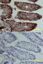
- Osteoprotegerin/TNFRSF11B in Mouse Intestine. Osteoprotegerin/TNFRSF11B was detected in perfusion fixed frozen sections of mouse intestine using Goat Anti-Mouse Osteoprotegerin/TNFRSF11B Biotinylated Antigen Affinity-purified Polyclonal Antibody (Catalog # BAF459) at 15 µg/mL overnight at 4 °C. Tissue was stained using the Anti-Goat HRP-DAB Cell & Tissue Staining Kit (brown; Catalog # CTS008) and counterstained with hematoxylin (blue). Lower panel shows a lack of labeling if primary antibodies are omitted and tissue is stained only with secondary antibody followed by incubation with detection reagents. View our protocol for Chromogenic IHC Staining of Frozen Tissue Sections.
