Explore
 Validate
 Learn
Learn Western blot
Antibody data
- Antibody Data
- Antigen structure
- References [0]
- Comments [0]
- Validations
- Western blot [1]
- Flow cytometry [1]
Submit
Validation data
Reference
Comment
Report error
- Product number
- 10-6001-25 - Provider product page

- Provider
- ABEOMICS Inc.
- Product name
- Anti-OPG Antibody
- Antibody type
- Monoclonal
- Description
- OPG (Osteoprotegerin) belongs to the TNF receptor super-family and is implicated in bone remodeling and in the atherosclerotic process. This molecule acts as a decoy receptor for the RANKL (Receptor Activator of Nuclear Factor-KappaB Ligand), inhibiting binding of RANKL to its receptor, RANK. OPG acts as a soluble neutralizing receptor of TRAIL (TNF-Related Apoptosis-Inducing Ligand), an anti-inflammatory molecule with anti-atherosclerotic properties. Mostly implicated in bone remodeling, the RANK/RANKL/OPG axis is involved in immune and vascular system. OPG has been associated with increased risk of atherosclerotic disease in the general population. OPG has also been associated with increased pulse wave velocity, progression of arterial calcification and with mortality in both end-stage renal failure patients as well as the general population.
- Reactivity
- Human
- Host
- Mouse
- Conjugate
- Unconjugated
- Antigen sequence
A partial length recombinant protei
n   (a.a 15-206)  of OPG was used a
s the immunogen for this antibody.- Isotype
- IgG
- Antibody clone number
- ABM10D2
- Vial size
- 100 µg
- Concentration
- 0.5 mg/ml
- Storage
- Store the antibody at 4°C, stable for 6 months. For long-term storage, store at -20°C. Avoid repeat freez thawing
No comments: Submit comment
Supportive validation
- Submitted by
- ABEOMICS Inc. (provider)
- Main image

- Experimental details
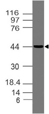
- Western blot analysis of OPG. Anti-OPG antibody (Clone: ABM10D2) was used at 4 µg/ml on Hela lysate.
- Protocol
- Protocol
Supportive validation
- Submitted by
- ABEOMICS Inc. (provider)
- Main image
- Experimental details
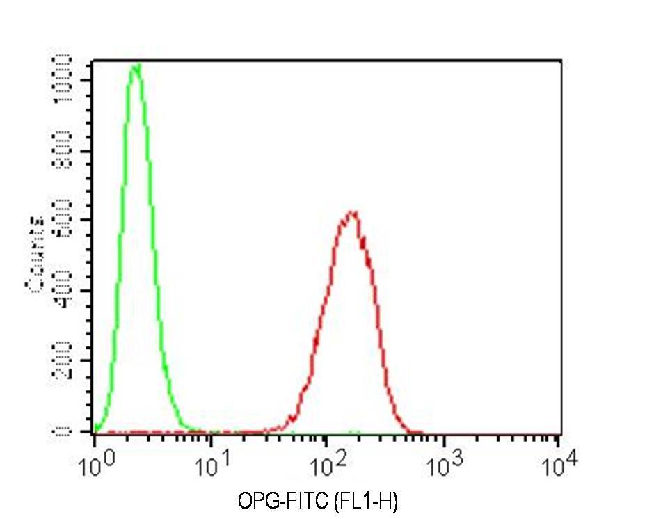
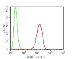
- Intracellular Flow analysis of OPG antibody on Ramos cells using 0.5 µg/ 10^6 cells of anti-OPG antibody (ABM10D2). Green represents isotype control; red represents anti-OPG antibody. Goat anti-mouse FITC conjugate was used as secondary antibody. (Cells were fixed with 4% paraformaldehyde for 10 min and washed with PBS by centrifuging at 1100 for 5 min followed by permeabilization for 20 min and washed again as mentioned above. Then cell were incubated with primary antibody for 45 min. and after washing the cells twice in PBS, incubated with conjugated secondary antibody for 30 min. Data acquisition was done after washing twice with PBS as mentioned above).
- Protocol
- Protocol