Explore
 Validate
 Learn
Learn Western blot
Antibody data
- Antibody Data
- Antigen structure
- References [0]
- Comments [0]
- Validations
- Western blot [3]
- Immunocytochemistry [1]
Submit
Validation data
Reference
Comment
Report error
- Product number
- PA5-17458 - Provider product page

- Provider
- Invitrogen Antibodies
- Product name
- DJ-1 Polyclonal Antibody
- Antibody type
- Polyclonal
- Antigen
- Synthetic peptide
- Description
- It is not recommended to aliquot this antibody.
- Reactivity
- Human, Mouse, Rat
- Host
- Rabbit
- Isotype
- IgG
- Vial size
- 100 μL
- Concentration
- 21 μg/mL
- Storage
- -20°C
No comments: Submit comment
Supportive validation
- Submitted by
- Invitrogen Antibodies (provider)
- Main image
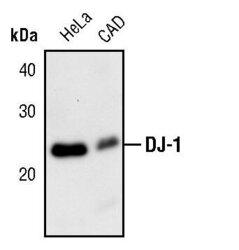
- Experimental details
- Western blot analysis of DJ-1 in extracts from HeLa and CAD cells using DJ-1 polyclonal antibody (Product # PA5-17458).

- Submitted by
- Invitrogen Antibodies (provider)
- Main image
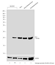
- Experimental details
- Western blot was performed using DJ-1 Polyclonal Antibody (Product # PA5-17458) and a 20 kDa band corresponding to DJ-1 was observed in HeLa, Mouse Skeletal Muscle and Rat Skeletal Muscle and increased upon Hydrogen peroxide treatment in SH-SY5Y. Whole cell extracts (30 µg lysate) of SH-SY5Y (Lane 1), SH-SY5Y treated with Hydrogen peroxide (25 mM for 24 hours) (Lane 2), HeLa (Lane 3) and tissue extracts (30 µg lysate) of Mouse Skeletal Muscle (Lane 4) and Rat Skeletal Muscle (Lane 5) were electrophoresed using Novex® NuPAGE® 12 % Bis-Tris gel (Product # NP0342BOX). Resolved proteins were then transferred onto a nitrocellulose membrane (Product # IB23001) by iBlot® 2 Dry Blotting System (Product # IB21001). The blot was probed with the primary antibody (1:1000 dilution) and detected by chemiluminescence with Goat anti-Rabbit IgG (Heavy Chain), Superclonal™ Recombinant Secondary Antibody, HRP (Product # A27036, 1:4000 dilution) using the iBright FL 1000 (Product # A32752). Chemiluminescent detection was performed using Novex® ECL Chemiluminescent Substrate Reagent Kit (Product # WP20005).
- Submitted by
- Invitrogen Antibodies (provider)
- Main image

- Experimental details
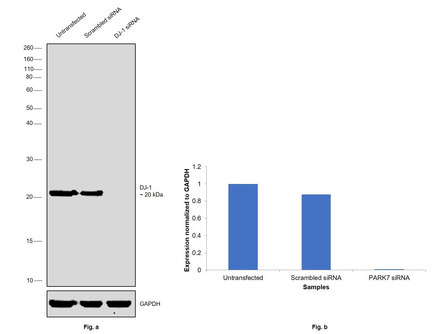
- Knockdown of DJ-1 was achieved by transfecting HeLa cells with DJ-1 specific siRNAs (Silencer® select Product # s22304). Western blot analysis (Fig. a) was performed using whole cell extracts from the DJ-1 knockdown cells (lane 3), non-specific scrambled siRNA transfected cells (lane 2) and untransfected cells (lane 1). The blot was probed with DJ-1 Polyclonal Antibody (Product # PA5-17458, 1:1000 dilution) and Goat anti-Rabbit IgG (Heavy Chain), Superclonal™ Recombinant Secondary Antibody, HRP (Product # A27036, 0.25 µg/mL, 1:4000 dilution). Densitometric analysis of this western blot is shown in histogram (Fig. b). Decrease in signal upon siRNA mediated knock down confirms that antibody is specific to DJ-1.
Supportive validation
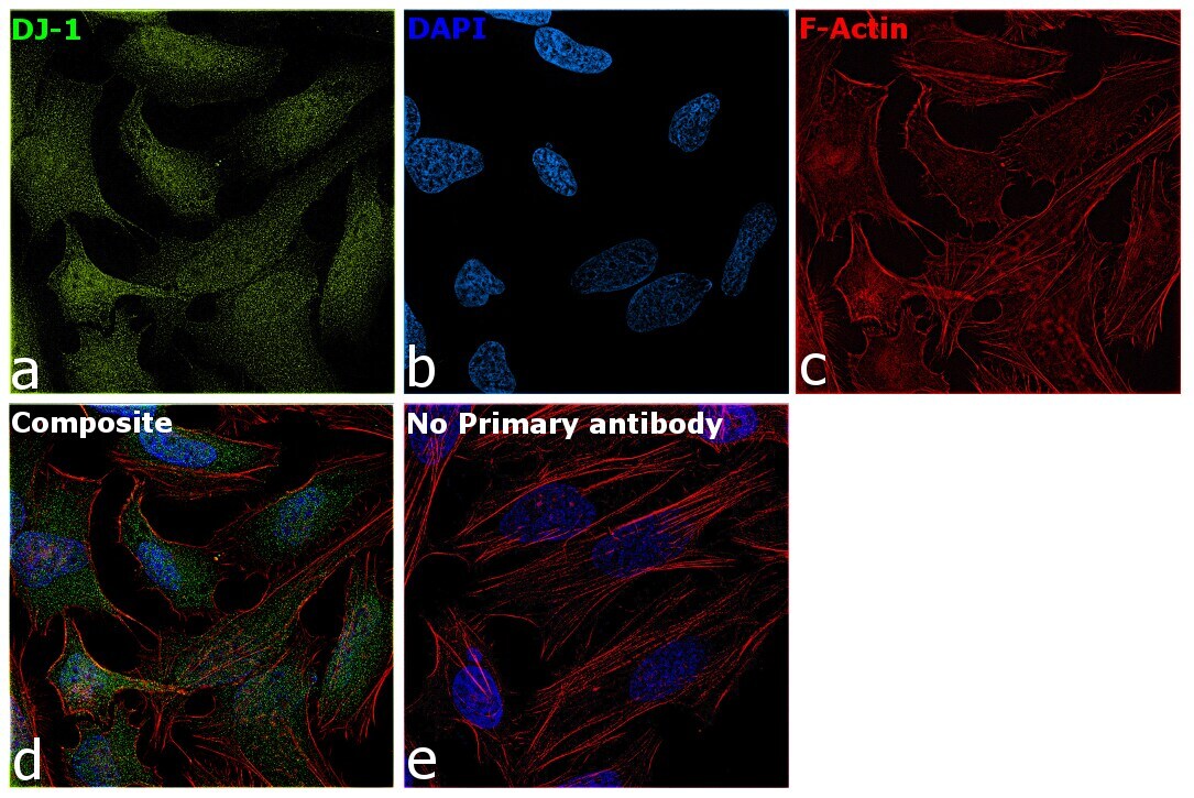
- Submitted by
- Invitrogen Antibodies (provider)
- Main image
- Experimental details
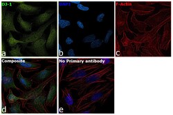
- Immunofluorescence analysis of DJ-1 was performed using 70% confluent log phase HeLa cells. The cells were fixed with 4% Paraformaldehyde for 10 minutes, permeabilized with 0.1% Triton™ X-100 for 10 minutes, and blocked with 2% BSA for 1 hour at room temperature. The cells were labeled with DJ-1 Polyclonal Antibody (Product # PA5-17458) at 1:200 dilution in 0.1% BSA, incubated at 4 degree Celsius overnight and then labeled with Donkey anti-Rabbit IgG (H+L) Highly Cross-Adsorbed Secondary Antibody, Alexa Fluor Plus 488 (Product # A32790, 1:2000 dilution) for 45 minutes at room temperature (Panel a: Green). Nuclei (Panel b: Blue) were stained with SlowFade® Gold Antifade Mountant with DAPI (Product # S36938). F-actin (Panel c: Red) was stained with Rhodamine Phalloidin (Product # R415, 1:300). Panel d represents the merged image showing cytoplasmic and nuclear localization. Panel e represents control cells with no primary antibody to assess background. The images were captured at 60X magnification.