Explore
 Validate
 Learn
Learn Western blot
Antibody data
- Antibody Data
- Antigen structure
- References [1]
- Comments [0]
- Validations
- Western blot [6]
- Immunocytochemistry [1]
- Immunohistochemistry [2]
Submit
Validation data
Reference
Comment
Report error
- Product number
- PA1-46262 - Provider product page

- Provider
- Invitrogen Antibodies
- Product name
- DJ-1 Polyclonal Antibody
- Antibody type
- Polyclonal
- Antigen
- Other
- Description
- This antibody is predicted to react with rat, bovine, zebra fish and Golden Syrian hamster based on 100% sequence homology.
- Concentration
- Conc. Not Determined
Submitted references Deficiency of Parkinson's Related Protein DJ-1 Alters Cdk5 Signalling and Induces Neuronal Death by Aberrant Cell Cycle Re-entry.
López-Grueso MJ, Padilla CA, Bárcena JA, Requejo-Aguilar R
Cellular and molecular neurobiology 2023 Mar;43(2):757-769
Cellular and molecular neurobiology 2023 Mar;43(2):757-769
No comments: Submit comment
Supportive validation
- Submitted by
- Invitrogen Antibodies (provider)
- Main image

- Experimental details
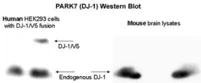
- Cells were transfected with the pCMV6-ENTRY control (lane 1) or pCMV6-ENTRY PARK7 cDNA (lane 2) for 48 hrs and lysed. Equivalent amounts of cell lysates (5 µg per lane) were separated by SDS-PAGE and immunoblotted with anti-PARK7.

- Submitted by
- Invitrogen Antibodies (provider)
- Main image

- Experimental details
- Western blot analysis of DJ-1 in 5 µg /lane cells transfected with the pCMV6-ENTRY. Samples were incubated in DJ-1 polyclonal antibody (Product # PA1-46262). Lane 1: control; Lane 2: pCMV6-ENTRY PARK7 cDNA. Incubated for 48 hrs and lysed. Equivalent amounts of cell lysates were separated by SDS-PAGE and immunoblotted with anti-Park7(DJ-1).
- Submitted by
- Invitrogen Antibodies (provider)
- Main image

- Experimental details
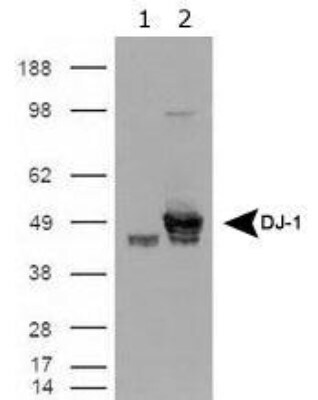
- Western blot analysis of DJ-1 in Human HEK293 cells and mouse brain lysates. Sample was incubated in DJ-1 polyclonal antibody (Product # PA1-46262).
- Submitted by
- Invitrogen Antibodies (provider)
- Main image

- Experimental details
- Western blot analysis of DJ-1 in 0.05 mg/mL Human Brain lysate. Samples were incubated in DJ-1 polyclonal antibody (Product # PA1-46262). This experiment was performed under reducing conditions using the 12-230 kDa separation system.

- Submitted by
- Invitrogen Antibodies (provider)
- Main image
- Experimental details
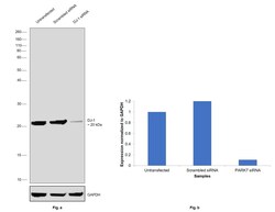
- Knockdown of DJ-1 was achieved by transfecting HeLa cells with DJ-1 specific siRNAs (Silencer® select Product # s22304). Western blot analysis (Fig. a) was performed using whole cell extracts from the DJ-1 knockdown cells (lane 3), non-specific scrambled siRNA transfected cells (lane 2) and untransfected cells (lane 1). The blot was probed with DJ-1 Polyclonal Antibody (Product # PA1-46262, 1:2000 dilution) and Goat anti-Rabbit IgG (H+L), Superclonal™ Recombinant Secondary Antibody, HRP (Product # A27036, 0.25 µg/mL, 1:4000 dilution). Densitometric analysis of this western blot is shown in histogram (Fig. b). Decrease in signal upon siRNA mediated knock down confirms that antibody is specific to DJ-1.

- Submitted by
- Invitrogen Antibodies (provider)
- Main image
- Experimental details
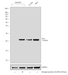
- Western blot was performed using DJ-1 Polyclonal Antibody (Product # PA1-46262) and a 20 kDa band corresponding to DJ-1 was observed in U-2 OS, HeLa and increased upon Hydrogen peroxide treatment in SH-SY5Y. Whole cell extracts (30 µg lysate) of SH-SY5Y (Lane 1), SH-SY5Y treated with Hydrogen peroxide (25 mM for 24 hours) (Lane 2), U-2 OS (Lane 3) and HeLa (Lane 4) were electrophoresed using Novex® NuPAGE® 12 % Bis-Tris gel (Product # NP0342BOX). Resolved proteins were then transferred onto a nitrocellulose membrane (Product # IB23001) by iBlot® 2 Dry Blotting System (Product # IB21001). The blot was probed with the primary antibody (1:2000 dilution) and detected by chemiluminescence with Goat anti-Rabbit IgG (H+L), Superclonal™ Recombinant Secondary Antibody, HRP (Product # A27036, 1:4000 dilution) using the iBright FL 1000 (Product # A32752). Chemiluminescent detection was performed using Novex® ECL Chemiluminescent Substrate Reagent Kit (Product # WP20005).
Supportive validation
- Submitted by
- Invitrogen Antibodies (provider)
- Main image
- Experimental details
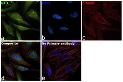
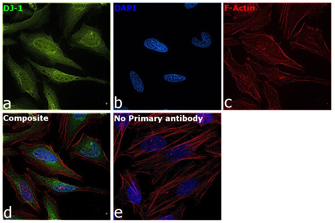
- Immunofluorescence analysis of DJ-1 was performed using 70% confluent log phase He-La cells. The cells were fixed with 4% Paraformaldehyde for 10 minutes, permeabilized with 0.1% Triton™ X-100 for 10 minutes, and blocked with 2% BSA for 1 hour at room temperature. The cells were labeled with DJ-1 Polyclonal Antibody (Product # PA1-46262) at 1:500 dilution in 0.1% BSA, incubated at 4 degree Celsius overnight and then labeled with Donkey anti-Rabbit IgG (H+L) Highly Cross-Adsorbed Secondary Antibody, Alexa Fluor Plus 488 (Product # A32790, 1:2000 dilution) for 45 minutes at room temperature (Panel a: Green). Nuclei (Panel b: Blue) were stained with SlowFade® Gold Antifade Mountant with DAPI (Product # S36938). F-actin (Panel c: Red) was stained with Rhodamine Phalloidin (Product # R415, 1:300). Panel d represents the merged image showing cytoplasmic and nuclear localization. Panel e represents control cells with no primary antibody to assess background. The images were captured at 60X magnification.
Supportive validation
- Submitted by
- Invitrogen Antibodies (provider)
- Main image

- Experimental details
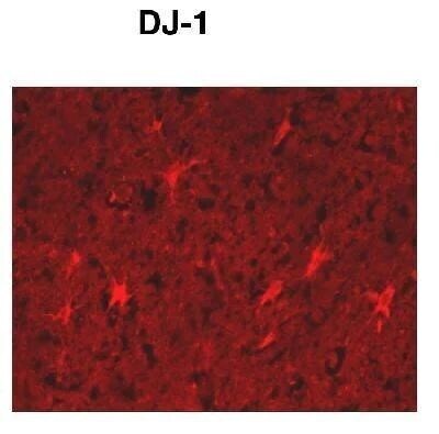
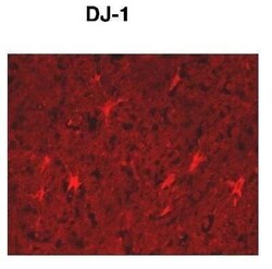
- Immunohistochemical analysis of DJ-1 in paraffin embedded human cortex. Samples were incubated in DJ-1 polyclonal antibody (Product # PA1-46262).

- Submitted by
- Invitrogen Antibodies (provider)
- Main image
- Experimental details
- Immunohistochemical analysis of DJ-1 in paraffin embedded human cortex. Samples were incubated in DJ-1 polyclonal antibody (Product # PA1-46262).