Explore
 Validate
 Learn
Learn Western blot
Antibody data
- Antibody Data
- Antigen structure
- References [1]
- Comments [0]
- Validations
- Western blot [2]
- Immunocytochemistry [1]
- Other assay [1]
Submit
Validation data
Reference
Comment
Report error
- Product number
- PA5-34739 - Provider product page

- Provider
- Invitrogen Antibodies
- Product name
- PDGFRA Polyclonal Antibody
- Antibody type
- Polyclonal
- Antigen
- Recombinant protein fragment
- Description
- Recommended positive controls: K562, PC-12. Predicted reactivity: Human (99%), Mouse (91%), Rat (92%), Xenopus laevis (82%), Chicken (86%), Bovine (96%). Store product as a concentrated solution. Centrifuge briefly prior to opening the vial.
- Reactivity
- Human, Mouse, Rat
- Host
- Rabbit
- Isotype
- IgG
- Vial size
- 100 µL
- Concentration
- 0.79 mg/mL
- Storage
- Store at 4°C short term. For long term storage, store at -20°C, avoiding freeze/thaw cycles.
Submitted references Knockdown of CD146 promotes endothelial-to-mesenchymal transition via Wnt/β-catenin pathway.
Zhang ZY, Zhai C, Yang XY, Li HB, Wu LL, Li L
PloS one 2022;17(8):e0273542
PloS one 2022;17(8):e0273542
No comments: Submit comment
Supportive validation
- Submitted by
- Invitrogen Antibodies (provider)
- Main image

- Experimental details
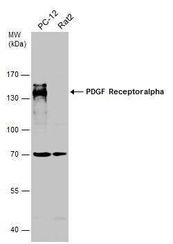
- Western Blot using PDGFRA Polyclonal Antibody (Product # PA5-34739). Sample (30 µg of whole cell lysate). A: K562 .5% SDS PAGE. PDGFRA Polyclonal Antibody (Product # PA5-34739) diluted at 1:1,000.
- Submitted by
- Invitrogen Antibodies (provider)
- Main image

- Experimental details
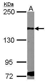
- Western Blot analysis of PDGFRA was performed by separating 30 µg of various whole cell extracts by 7.5% SDS-PAGE. Proteins were transferred to a membrane and probed with a PDGFRA Polyclonal Antibody (Product # PA5-34739) at a dilution of 1:500.
Supportive validation
- Submitted by
- Invitrogen Antibodies (provider)
- Main image

- Experimental details
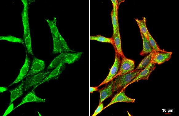
- PDGFRA Polyclonal Antibody detects PDGF Receptor alpha protein at cytoplasm by immunofluorescent analysis. Sample: NIH/3T3 cells were fixed in 4% paraformaldehyde at RT for 15 min. Green: PDGF Receptor alpha stained by PDGFRA Polyclonal Antibody (Product # PA5-34739) diluted at 1:500. Red: phalloidin, a cytoskeleton marker diluted at 1:200. Blue: Hoechst 33342 staining.
Supportive validation
- Submitted by
- Invitrogen Antibodies (provider)
- Main image
- Experimental details
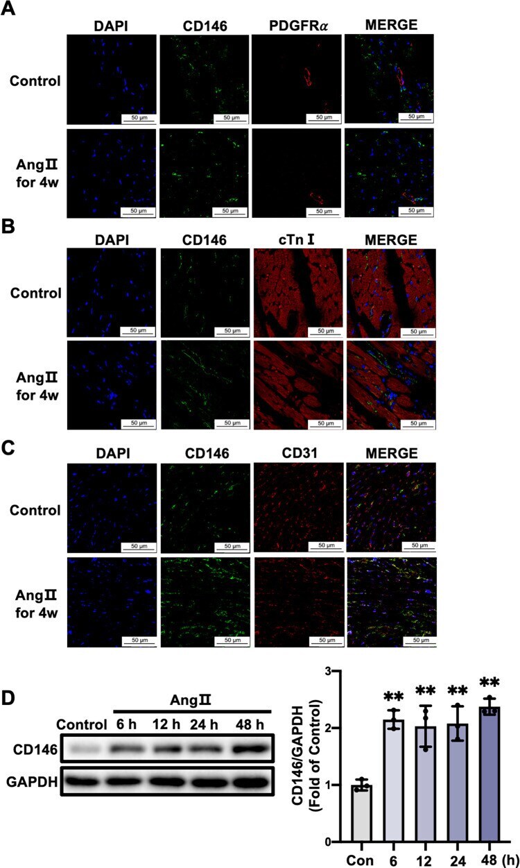
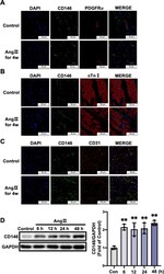
- 10.1371/journal.pone.0273542.g003 Fig 3 CD146 upregulation by Ang II is mainly restricted to cardiac ECs. Representative double-immunofluorescence images of CD146 with PDGFRalpha ( A ), cTnI ( B ) and CD31 ( C ) in left ventricles from control and 4-week Ang II-infused mice (scale bars indicate 50 mum). Green represents CD146; red represents PDGFRalpha, cTnI and CD31, respectively; blue represents nuclei. D HUVECs were treated with Ang II (1x10 -6 mol/L) for the indicated times and the protein levels of CD146 were measured by Western blot analysis. GAPDH served as an internal control. * P