Explore
 Validate
 Learn
Learn Western blot
 Immunocytochemistry
Antibody data
- Antibody Data
- Antigen structure
- References [5]
- Comments [0]
- Validations
- Immunocytochemistry [1]
- Immunohistochemistry [1]
Submit
Validation data
Reference
Comment
Report error
- Product number
- HPA004426 - Provider product page

- Provider
- Atlas Antibodies
- Proper citation
- Atlas Antibodies Cat#HPA004426, RRID:AB_1079701
- Product name
- Anti-PSAP
- Antibody type
- Polyclonal
- Description
- Polyclonal Antibody against Human PSAP, Gene description: prosaposin, Alternative Gene Names: GLBA, SAP1, Validated applications: ICC, IHC, WB, Uniprot ID: P07602, Storage: Store at +4°C for short term storage. Long time storage is recommended at -20°C.
- Reactivity
- Human
- Host
- Rabbit
- Conjugate
- Unconjugated
- Isotype
- IgG
- Vial size
- 100 µl
- Concentration
- 0.1 mg/ml
- Storage
- Store at +4°C for short term storage. Long time storage is recommended at -20°C.
- Handling
- The antibody solution should be gently mixed before use.
Submitted references The exocyst complex is an essential component of the mammalian constitutive secretory pathway
Prosaposin maintains lipid homeostasis in dopamine neurons and counteracts experimental parkinsonism in rodents
Decreased Prosaposin and Progranulin in the Cingulate Cortex Are Associated with Schizophrenia Pathophysiology.
Prosaposin Reduces α-Synuclein in Cells and Saposin C Dislodges it from Glucosylceramide-enriched Lipid Membranes.
Progranulin mutations result in impaired processing of prosaposin and reduced glucocerebrosidase activity
Pereira C, Stalder D, Anderson G, Shun-Shion A, Houghton J, Antrobus R, Chapman M, Fazakerley D, Gershlick D
Journal of Cell Biology 2023;222(5)
Journal of Cell Biology 2023;222(5)
Prosaposin maintains lipid homeostasis in dopamine neurons and counteracts experimental parkinsonism in rodents
He Y, Kaya I, Shariatgorji R, Lundkvist J, Wahlberg L, Nilsson A, Mamula D, Kehr J, Zareba-Paslawska J, Biverstål H, Chergui K, Zhang X, Andren P, Svenningsson P
Nature Communications 2023;14(1)
Nature Communications 2023;14(1)
Decreased Prosaposin and Progranulin in the Cingulate Cortex Are Associated with Schizophrenia Pathophysiology.
He Y, Zhang X, Flais I, Svenningsson P
International journal of molecular sciences 2022 Oct 10;23(19)
International journal of molecular sciences 2022 Oct 10;23(19)
Prosaposin Reduces α-Synuclein in Cells and Saposin C Dislodges it from Glucosylceramide-enriched Lipid Membranes.
Kojima R, Zurbruegg M, Li T, Paslawski W, Zhang X, Svenningsson P
Journal of molecular neuroscience : MN 2022 Nov;72(11):2313-2325
Journal of molecular neuroscience : MN 2022 Nov;72(11):2313-2325
Progranulin mutations result in impaired processing of prosaposin and reduced glucocerebrosidase activity
Krainc D, Zheng J, Young T, Ysselstein D, Valdez C
Human Molecular Genetics 2020;29(5):716-726
Human Molecular Genetics 2020;29(5):716-726
No comments: Submit comment
Supportive validation
- Submitted by
- Atlas Antibodies (provider)
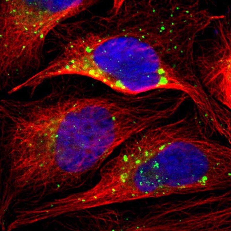
- Main image

- Experimental details
- Immunofluorescent staining of human cell line U-2 OS shows localization to vesicles.
- Sample type
- Human
Supportive validation
- Submitted by
- Atlas Antibodies (provider)
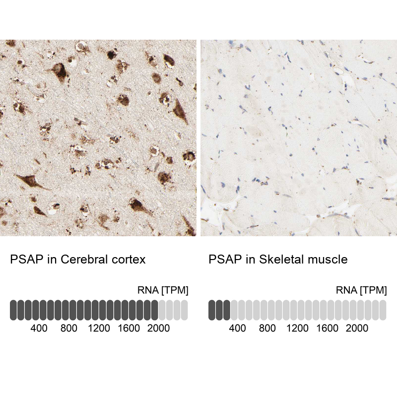
- Enhanced method
- Orthogonal validation
- Main image
- Experimental details
- Immunohistochemistry analysis in human cerebral cortex and skeletal muscle tissues using HPA004426 antibody. Corresponding PSAP RNA-seq data are presented for the same tissues.
- Sample type
- Human
- Protocol
- Protocol