Explore
 Validate
 Learn
Learn Western blot
 Immunocytochemistry
Antibody data
- Antibody Data
- Antigen structure
- References [0]
- Comments [0]
- Validations
- Western blot [1]
- Immunohistochemistry [1]
Submit
Validation data
Reference
Comment
Report error
- Product number
- MAB6977 - Provider product page

- Provider
- Novus Biologicals
- Product name
- Mouse Monoclonal Zyxin Antibody
- Antibody type
- Monoclonal
- Description
- Protein A or G purified from hybridoma culture supernatant. Detects human Zyxin in direct ELISAs and Western blots.
- Reactivity
- Human, Mouse, Rat
- Host
- Mouse
- Conjugate
- Unconjugated
- Isotype
- IgG
- Vial size
- 100 ug
- Concentration
- LYOPH
- Storage
- Use a manual defrost freezer and avoid repeated freeze-thaw cycles. 12 months from date of receipt, -20 to -70 degreesC as supplied. 1 month, 2 to 8 degreesC under sterile conditions after reconstitution. 6 months, -20 to -70 degreesC under sterile conditions after reconstitution.
No comments: Submit comment
Supportive validation
- Submitted by
- Novus Biologicals (provider)
- Main image

- Experimental details
- Detection of Human, Mouse, and Rat Zyxin by Western Blot. Western blot shows lysates of Human cervical epithelial carcinoma cell line, PT18 mouse mast/ basophil cell line, Neuro-2A mouse neuroblastoma cell line, L6 rat myoblast cell line, and PC-12 rat adrenal pheochromocytoma cell line. PVDF membrane was probed with 0.5 µg/mL of Mouse Anti-Human/Mouse/Rat Zyxin Monoclonal Antibody (Catalog # MAB6977) followed by HRP-conjugated Anti-Mouse IgG Secondary Antibody (Catalog # HAF007). A specific band was detected for Zyxin at approximately 82 kDa (as indicated). This experiment was conducted under reducing conditions and using Immunoblot Buffer Group 1.

Supportive validation
- Submitted by
- Novus Biologicals (provider)
- Main image

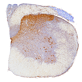
- Experimental details
- Zyxin in Rat Spinal Cord. Zyxin was detected in perfusion fixed frozen sections of rat spinal cord using Mouse Anti-Human/Mouse/Rat Zyxin Monoclonal Antibody (Catalog # MAB6977) at 3 µg/mL for 1 hour at room temperature followed by incubation with the Anti-Mouse IgG VisUCyte™ HRP Polymer Antibody (Catalog # VC001). Tissue was stained using DAB (brown) and counterstained with hematoxylin (blue). Specific staining was localized to motor neurons and dorsal horn. View our protocol for IHC Staining with VisUCyte HRP Polymer Detection Reagents.