 Explore
 Validate
 Learn
Learn Western blot
 ELISA
ELISAAntibody data
- Antibody Data
- Antigen structure
- References [0]
- Comments [0]
- Validations
- Western blot [3]
- Immunohistochemistry [1]
- Other assay [1]
Submit
Validation data
Reference
Comment
Report error
- Product number
- MA1-187 - Provider product page

- Provider
- Invitrogen Antibodies
- Product name
- Progranulin Monoclonal Antibody (2D4-2F1)
- Antibody type
- Monoclonal
- Antigen
- Recombinant full-length protein
- Description
- By Western blot, MA1-187 detects Human Progranulin at ~85 kDa.
- Reactivity
- Human
- Host
- Mouse
- Isotype
- IgG
- Antibody clone number
- 2D4-2F1
- Vial size
- 200 µL
- Concentration
- 1.0 mg/mL
- Storage
- -20°C
No comments: Submit comment
Supportive validation
- Submitted by
- Invitrogen Antibodies (provider)
- Main image

- Experimental details
- Western blot analysis of recombinant Progranulin was performed by loading the indicated amounts of protein, and 15 µL of PageRuler Prestained Protein Ladder (Product # 26616) per well onto a 4-20% Tris-HCl polyacrylamide gel. Proteins were transferred to a PVDF membrane using the G2 Fast Blotter (Product # 62288) and blocked with StartingBlock (PBS) Blocking Buffer (Product # 37538) for at least 1 hour at room temperature. Recombinant protein was detected at ~85 kD using a anti Progranulin monoclonal antibody (Product # MA1-187) at a concentration of 4 µg/mL in blocking buffer overnight at 4C on a rocking platform, followed by an HRP-conjugated goat anti-mouse IgG (light chain specific) secondary antibody at a dilution of 1:10,000 for at least 1 hour. Chemiluminescent detection was performed using SuperSignal West Dura (Product # 34075).

- Submitted by
- Invitrogen Antibodies (provider)
- Main image

- Experimental details
- Western blot analysis of Progranulin was performed by loading 40 µg of the indicated whole cell lysates per well, and 15 µL of PageRuler Prestained Protein Ladder (Product # 26616) per well onto a 4-20% Tris-HCl polyacrylamide gel. Proteins were transferred to a PVDF membrane using the G2 Fast Blotter (Product # 62288) and blocked with StartingBlock (PBS) Blocking Buffer (Product # 37538) for at least 1 hour at room temperature. Progranulin was detected at ~85 kD using a anti Progranulin monoclonal antibody (Product # MA1-187) at a concentration of 4 µg/mL in blocking buffer overnight at 4C on a rocking platform, followed by an HRP-conjugated goat anti-mouse IgG (light chainspecific) secondary antibody at a dilution of 1:10,000 for at least 1 hour. Chemiluminescent detection was performed using SuperSignal West Dura (Product # 34075).

- Submitted by
- Invitrogen Antibodies (provider)
- Main image

- Experimental details
- Western blot of Progranulin was performed by loading 50 µg of WT (lane 1) and GRN CRISPR KO (lane 2) HEK293T cell lysates in RIPA buffer onto a 5-16% gradient polyacrylamide gel. Proteins on the blots were visualized with Ponceau staining (below immunoblot). Proteins were transferred to nitrocellulose membrane and blocked in 5% milk for 1 hr. GRN was detected at approximately 64 kDa using a GRN monoclonal antibody (Product # MA1-187) at a dilution of 1:5,000 in 5% BSA in TBS with 0.1% Tween 20 (TBST) overnight at 4°C. The peroxidase-conjugated secondary antibody (Product # 62-6520) was diluted to 0.2 µg/mL in TBST with 5% milk for 1 hr. Chemiluminescent detection was performed using Pierce ECL Western Blotting Substrate (Product # 32106). Data courtesy of YCharOS Inc., an open science company with the mission of characterizing commercially available antibodies using knockout validation.

Supportive validation
- Submitted by
- Invitrogen Antibodies (provider)
- Main image
- Experimental details
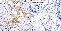
- Immunohistochemistry analysis of progranulin was performed on human kidney tissue and is showing positive staining (left panel). To expose target proteins, antigen retrieval was performed by microwaving tissues for 15 minutes in 10mM sodium citrate buffer (pH 6.0). Tissue slides were probed with progranulin monoclonal antibody (Product # MA1-187) at a dilution of 1:20, over night at 4C in a humidified chamber without (right panel) or with progranulin antibody (left panel). Tissues were washed, and incubated with secondary antibody (conjugated with HRP) for 30 min at room temperature in a humidified chamber. Detection was performed using a DAB substrate kit. Tissues were counterstained with hematoxylin and dehydrated to prep for mounting. Images were taken at 20x magnification.

Supportive validation
- Submitted by
- Invitrogen Antibodies (provider)
- Main image

- Experimental details
- Sandwich ELISA analysis of serum progranulin was performed by coating wells of a plate with monoclonal capture antibody at a concentration of 1.5 µg/mL overnight at 4C. The plate was washed 3 times with ELISA Wash Buffer (Product # N503) and blocked with blocking buffer (Product # 37538) for one hour at room temperature. Human sepsis serum sample was initially diluted 1:4 and further serially diluted 1:2 for 11 dilutions. 100 µL of diluted serum samples were added to wells in duplicate and incubated for 2 hours at room temperature. The plate was washed, and then incubated with 100 µL per well of Biotinylated mouse anti Progranulin antibody (Product # MA1-187) at a concentration of 3 µg/mL for 1 hour at room temperature. The plate was washed and incubated with conjugated Streptavidin-HRP (Product # 21130) at a dilution of 1:10,000 for 1 hour at room temperature, and washed again with ELISA Wash Buffer. The plate was developed by incubating 100 µL per well of 1-Step Ultra TMB substrate (Product # 34028) per well for 20 minutes at room temperature in the dark. The reaction was stopped with 100 µL per well of Stop solution (Product # N600). Absorbances were read on a spectrophotometer at 450-550 nm.
