Explore
 Validate
 Learn
Learn Western blot
 Immunocytochemistry
Antibody data
- Antibody Data
- Antigen structure
- References [2]
- Comments [0]
- Validations
- Immunocytochemistry [1]
- Immunohistochemistry [1]
- Other assay [4]
Submit
Validation data
Reference
Comment
Report error
- Product number
- PA5-27275 - Provider product page

- Provider
- Invitrogen Antibodies
- Product name
- Granulins Polyclonal Antibody
- Antibody type
- Polyclonal
- Antigen
- Recombinant full-length protein
- Description
- Recommended positive controls: HepG2, mouse kidney, rat kidney. Store product as a concentrated solution. Centrifuge briefly prior to opening the vial.
- Reactivity
- Human
- Host
- Rabbit
- Isotype
- IgG
- Vial size
- 100 μL
- Concentration
- 1.43 mg/mL
- Storage
- Store at 4°C short term. For long term storage, store at -20°C, avoiding freeze/thaw cycles.
Submitted references Progranulin sustains STAT3 hyper-activation and oncogenic function in colorectal cancer cells.
Granulin, a novel STAT3-interacting protein, enhances STAT3 transcriptional function and correlates with poorer prognosis in breast cancer.
Laudisi F, Cherubini F, Di Grazia A, Dinallo V, Di Fusco D, Franzè E, Ortenzi A, Salvatori I, Scaricamazza S, Monteleone I, Sakamoto N, Monteleone G, Stolfi C
Molecular oncology 2019 Oct;13(10):2142-2159
Molecular oncology 2019 Oct;13(10):2142-2159
Granulin, a novel STAT3-interacting protein, enhances STAT3 transcriptional function and correlates with poorer prognosis in breast cancer.
Yeh JE, Kreimer S, Walker SR, Emori MM, Krystal H, Richardson A, Ivanov AR, Frank DA
Genes & cancer 2015 Mar;6(3-4):153-68
Genes & cancer 2015 Mar;6(3-4):153-68
No comments: Submit comment
Supportive validation
- Submitted by
- Invitrogen Antibodies (provider)
- Main image
- Experimental details
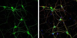
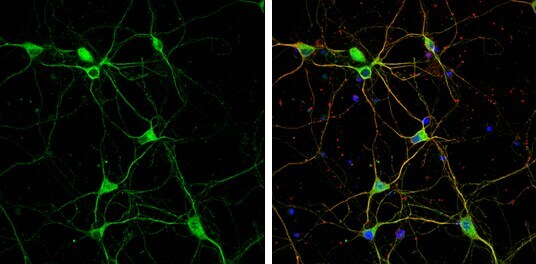
- Immunocytochemistry-Immunofluorescence analysis of Granulins was performed in DIV9 rat E18 primary cortical neuron cells fixed in 4% paraformaldehyde at RT for 15 min. Green: Granulins Polyclonal Antibody (Product # PA5-27275) diluted at 1:500. Red: beta Tubulin 3/ Tuj1, stained by beta Tubulin 3/ Tuj1 antibody. Blue: Fluoroshield with DAPI.
Supportive validation
- Submitted by
- Invitrogen Antibodies (provider)
- Main image
- Experimental details
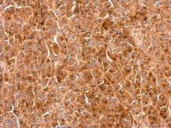
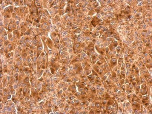
- Granulins Polyclonal Antibody detects GRN protein at cytosol on HBL435 xenograft by immunohistochemical analysis. Sample: Paraffin-embedded HBL435 xenograft. Granulins Polyclonal Antibody (Product # PA5-27275) dilution: 1:500. Antigen Retrieval: EDTA based buffer, pH 8.0, 15 min.
Supportive validation
- Submitted by
- Invitrogen Antibodies (provider)
- Main image

- Experimental details
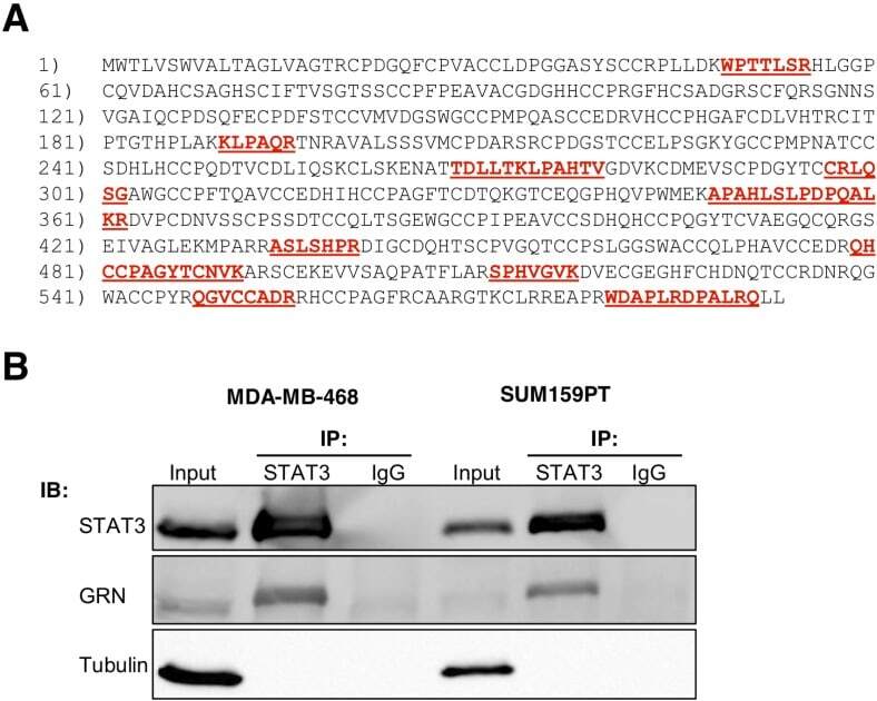
- Fig. 1 STAT3 interacts with GRN in breast cancer cells (A) Mass spectrometry coverage of GRN amino acid sequence P28799. Peptides identified by PEAKS or Proteome Discoverer are indicated in red underline. (B) STAT3-GRN interaction in MDA-MB-468 and SUM159PT cells was analyzed by immunoprecipitation using antibodies against STAT3 (or non-specific immunoglobulin G, IgG) followed by immunoblots with the indicated antibodies. Input indicates 5% of pre-immunoprecipitated samples.
- Submitted by
- Invitrogen Antibodies (provider)
- Main image
- Experimental details
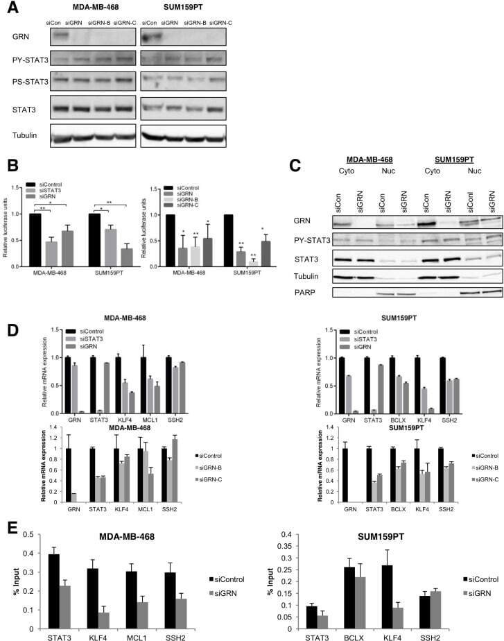
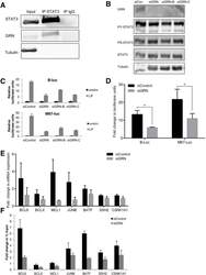
- Fig. 2 GRN is necessary for constitutive STAT3 transcriptional activity in triple-negative breast cancer cells MDA-MB-468 and SUM159PT cells were transfected with siRNA targeting GRN (pool siGRN and individual constructs siGRN-B and siGRN-C) or a non-targeting control (siCon). They were then analyzed by (A) immunoblot for levels of phosphorylated and total STAT3 in whole cell lysates, (B) luciferase reporter assay for STAT3-dependent transcriptional activity (N = 3), (C) nuclear and cytoplasmic fractions (tubulin serves as a loading control for the cytoplasmic fraction and PARP serves as a loading control for the nuclear fraction), (D) qRT-PCR for expression of endogenous STAT3 target genes (normalized to GAPDH; N = 3), and (E) chromatin immunoprecipitation with an antibody for STAT3 followed by PCR at the indicated STAT3 target genes (representative of N = 3).
- Submitted by
- Invitrogen Antibodies (provider)
- Main image
- Experimental details
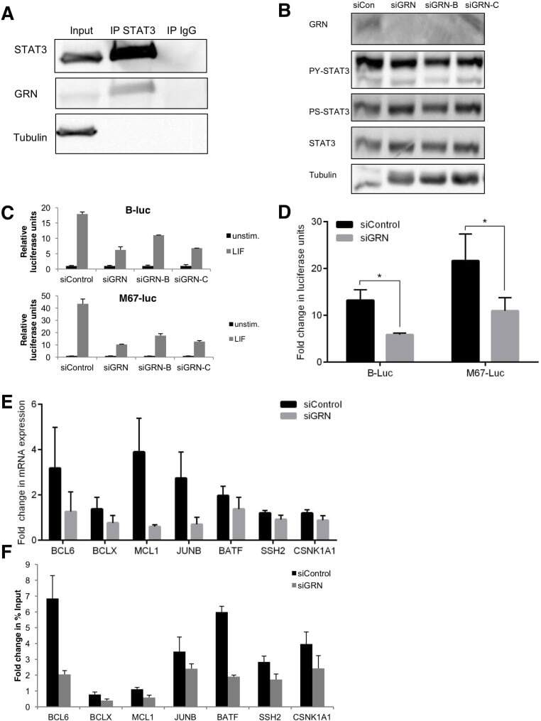
- Fig. 3 GRN is necessary for maximal cytokine-induced STAT3 transcriptional activity in breast cancercells (A) STAT3-GRN interaction in SK-BR-3 cells was analyzed by immunoprecipitation using antibodies against STAT3 (or non-specific IgG) followed by immunoblot with the indicated antibodies. Input indicates 5% of pre-immunoprecipitated samples. SK-BR-3 cells were transfected with siRNA targeting GRN (pool siGRN and individual constructs siGRN-B and siGRN-C) or a non-targeting control (siCon), then stimulated with LIF were analyzed by (B) immunoblot and (C) luciferase reporter assay for STAT3-dependent transcriptional activity. SK-BR-3 cells transfected with siRNA targeting GRN (pool) were analyzed by (D) luciferase reporter assay for STAT3-dependent transcriptional activity (B-luc, M67-luc) (N = 3), (E) qRT-PCR for expression of endogenous STAT3 target genes (normalized to HPRT; N = 6), and (F) chromatin immunoprecipitation with an antibody to STAT3 followed by PCR at the indicated STAT3 target genes (data normalized to unstimulated cells; representative of N = 2).
- Submitted by
- Invitrogen Antibodies (provider)
- Main image
- Experimental details
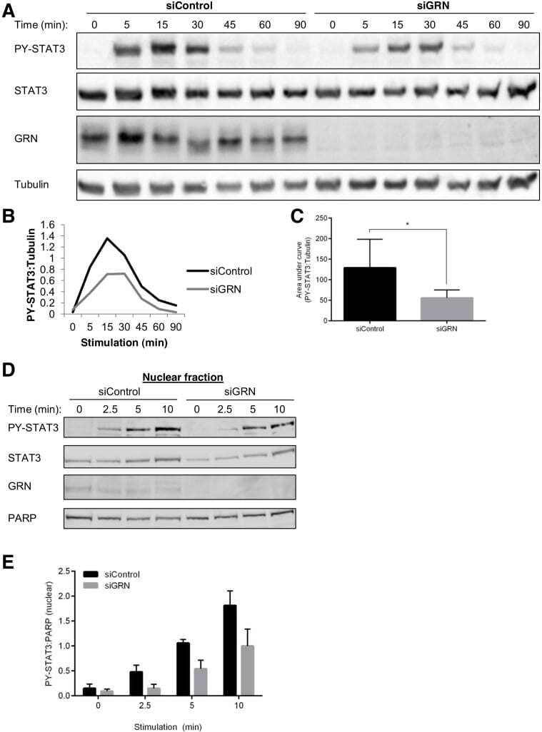
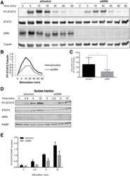
- Fig. 4 GRN enhances the kinetics of cytokine-stimulated STAT3 activation and nuclear translocation (A) SK-BR-3 cells were transfected with siRNA targeting GRN or a non-targeting control, then stimulated with LIF for the indicated times. Cells were then analyzed by immunoblot for levels of phosphorylated and total STAT3 in whole cell extracts. Tubulin serves as a loading control. (B) Immunoblot band intensities plotted as ratios of phosphorylated STAT3 to tubulin. (C) Area under the curve of phospho-STAT3:tubulin (N = 5). (D) SK-BR-3 cells were transfected with siRNA targeting GRN or a non-targeting control, then stimulated with LIF for the indicated times. Cells were then analyzed by immunoblot for levels of phosphorylated and total STAT3 in nuclear fractions. PARP serves as a nuclear fraction loading control. (E) Immunoblot band intensities plotted as ratios of phospho-STAT3 to PARP (N = 4).