 Explore
 Validate
 Learn
Learn Western blot
Antibody data
- Antibody Data
- Antigen structure
- References [0]
- Comments [0]
- Validations
- Western blot [1]
- Immunocytochemistry [1]
- Immunohistochemistry [6]
Submit
Validation data
Reference
Comment
Report error
- Product number
- AMAb91191 - Provider product page

- Provider
- Atlas Antibodies
- Proper citation
- Atlas Antibodies Cat#AMAb91191, RRID:AB_2665838
- Product name
- Anti-DAXX
- Antibody type
- Monoclonal
- Reactivity
- Human
- Host
- Mouse
- Conjugate
- Unconjugated
- Antigen sequence
ARGSSSSGGKKCYKLENEKLFEEFLELCKMQTADH
PEVVPFLYNRQQRAHSLFLASAEFCNILSRVLSRA
RSRPAKLYVYINELCTVLKAHSAKKKLNLAPAATT
SNEPSGNNPPTHLSLDPTNAENTASQSPRTR- Epitope
- Binds to an epitope located within the peptide sequence PEVVPFLYNR as determined by overlapping synthetic peptides.
- Isotype
- IgG
- Antibody clone number
- CL3580
- Vial size
- 100 µl
- Storage
- Store at +4°C for short term storage. Long time storage is recommended at -20°C.
No comments: Submit comment
Supportive validation
- Submitted by
- Atlas Antibodies (provider)
- Main image
- Experimental details
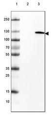
- Lane 1: Marker [kDa] 250, 130, 100, 70, 55, 35, 25, 15, 10Lane 2: Negative control (vector only transfected HEK293T lysate)Lane 3: Over-expression lysate (Co-expressed with a C-terminal myc-DDK tag (~3.1 kDa) in mammalian HEK293T cells, LY427987)

Supportive validation
- Submitted by
- Atlas Antibodies (provider)
- Main image

- Experimental details
- Immunofluorescence staining of HeLa cells using the Anti-DAXX monoclonal antibody, showing specific staining in the nucleoplasm and cytosol in green. Microtubule- and nuclear probes are visualized in red and blue, respectively (where available).
- Sample type
- HUMAN

Supportive validation
- Submitted by
- Atlas Antibodies (provider)
- Main image

- Experimental details
- Immunohistochemical staining of human cervix shows moderate nuclear immunoreactivity in epithelial and underlying connective tissue cells.

- Submitted by
- Atlas Antibodies (provider)
- Main image

- Experimental details
- Immunohistochemical staining of human breast shows nuclear positivity in glandular cells.

- Submitted by
- Atlas Antibodies (provider)
- Main image
- Experimental details
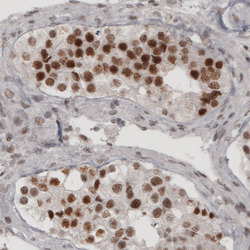
- Immunohistochemical staining of human testis shows nuclear immunoreactivity in seminiferous tubules cells.

- Submitted by
- Atlas Antibodies (provider)
- Main image
- Experimental details
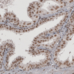
- Immunohistochemical staining of human prostate shows moderate nuclear positivity in glandular cells.

- Submitted by
- Atlas Antibodies (provider)
- Main image

- Experimental details
- Immunohistochemical staining of human tonsil shows nuclear immunoreactivity in lymphoid cells.

- Submitted by
- Atlas Antibodies (provider)
- Main image
- Experimental details
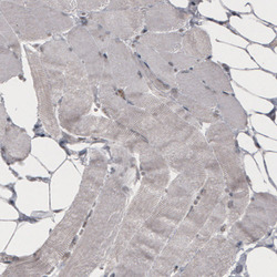
- Immunohistochemical staining of human skeletal muscle shows absence of immunoreactivity (negative control).
