Explore
 Validate
 Learn
Learn Western blot
 Immunohistochemistry
Antibody data
- Antibody Data
- Antigen structure
- References [0]
- Comments [0]
- Validations
- Western blot [1]
Submit
Validation data
Reference
Comment
Report error
- Product number
- PA1809 - Provider product page

- Provider
- Boster Biological Technology
- Product name
- Anti-Daxx Antibody
- Antibody type
- Polyclonal
- Description
- Polyclonal antibody for DAXX detection. Host: Rabbit.Size: 100μg/vial. Tested applications: WB, IHC-P, FCM. Reactive species: Human;Mouse;Rat. DAXX information: Molecular Weight: 81373 MW; Subcellular Localization: Cytoplasm. Nucleus, nucleoplasm. Nucleus, PML body . Nucleus, nucleolus. Chromosome, centromere. Dispersed throughout the nucleoplasm, in PML/POD/ND10 nuclear bodies, and in nucleoli. Colocalizes with histone H3.3, ATRX, HIRA and ASF1A at PML-nuclear bodies. Colocalizes with a subset of interphase centromeres, but is absent from mitotic centromeres. Detected in cytoplasmic punctate structures. Translocates from the nucleus to the cytoplasm upon glucose deprivation or oxidative stress. Colocalizes with RASSF1 in the nucleus. Colocalizes with USP7 in nucleoplasma with accumulation in speckled structures; Tissue Specificity: Ubiquitous.
- Reactivity
- Human, Mouse, Rat
- Host
- Rabbit
- Vial size
- 100μg/vial
- Concentration
- Add 0.2ml of distilled water will yield a concentration of 500ug/ml.
- Storage
- At -20°C for one year. After reconstitution, at 4°C for one month. It can also be aliquoted and stored frozen at -20°C for a longer time. Avoid repeated freezing and thawing.
- Handling
- Add 0.2ml of distilled water will yield a concentration of 500ug/ml.
No comments: Submit comment
Supportive validation
- Submitted by
- Boster Biological Technology (provider)
- Main image

- Experimental details
- Western blot analysis of DAXX using anti-DAXX antibody (PA1809). Electrophoresis was performed on a 5-20% SDS-PAGE gel at 70V (Stacking gel) / 90V (Resolving gel) for 2-3 hours. The sample well of each lane was loaded with 50ug of sample under reducing conditions. Lane 1: human Caco-2 whole cell lysates, Lane 2: human HepG2 whole cell lysates, Lane 3: human A549 whole cell lysates, Lane 4: human Raji whole cell lysates, Lane 5: rat testicular tissue lysates, Lane 6: mouse testicular tissue lysates. After Electrophoresis, proteins were transferred to a Nitrocellulose membrane at 150mA for 50-90 minutes. Blocked the membrane with 5% Non-fat Milk/ TBS for 1.5 hour at RT. The membrane was incubated with rabbit anti-DAXX antigen affinity purified polyclonal antibody (Catalog # PA1809) at 0.5 μg/mL overnight at 4°C, then washed with TBS-0.1%Tween 3 times with 5 minutes each and probed with a goat anti-rabbit IgG-HRP secondary antibody at a dilution of 1:10000 for 1.5 hour at RT. The signal is developed using an Enhanced Chemiluminescent detection (ECL) kit (Catalog # EK1002) with Tanon 5200 system. A specific band was detected for DAXX at approximately 110KD. The expected band size for DAXX is at 81KD.
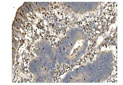
- Additional image