 Explore
 Validate
 Learn
Learn Western blot
Antibody data
- Antibody Data
- Antigen structure
- References [0]
- Comments [0]
- Validations
- Western blot [2]
- Immunocytochemistry [2]
- Immunohistochemistry [1]
Submit
Validation data
Reference
Comment
Report error
- Product number
- PA5-11720 - Provider product page

- Provider
- Invitrogen Antibodies
- Product name
- BMP-7 Polyclonal Antibody
- Antibody type
- Polyclonal
- Antigen
- Synthetic peptide
- Description
- This antibody is predicted to react with mouse based on sequence homology.
- Reactivity
- Human
- Host
- Rabbit
- Isotype
- IgG
- Vial size
- 400 µL
- Concentration
- 1.5 mg/mL
- Storage
- Store at 4°C short term. For long term storage, store at -20°C, avoiding freeze/thaw cycles.
No comments: Submit comment
Supportive validation
- Submitted by
- Invitrogen Antibodies (provider)
- Main image
- Experimental details
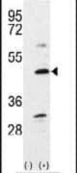
- Western blot analysis of Bmp7 (arrow) using a Bmp7 polyclonal antibody (Product # PA5-11720) in 293 cell lysates (2 µg/lane) either nontransfected (Lane 1) or transiently transfected with the Bmp7 gene (Lane 2).

- Submitted by
- Invitrogen Antibodies (provider)
- Main image

- Experimental details
- Western blot was performed using Anti-BMP-7 Polyclonal Antibody (Product # PA5-11720) and a 50 kDa band corresponding to Bmp7 was observed across cell lines tested. The expression was found to be low in HeLa, PC-3, HCT 116, BJ and Hep G2 as reported. Whole cell extracts (30 µg lysate) of MCF7 (Lane 1), T-47D (Lane 2), HeLa (Lane 3), PC-3 (Lane 4), HCT 116 (Lane 5), BJ (Lane 6) and Hep G2 (Lane 7) were electrophoresed using NuPAGE™ 4-12% Bis-Tris Protein Gel (Product # NP0321BOX), 10 well. Resolved proteins were then transferred onto a nitrocellulose membrane (Product # IB23001) by iBlot® 2 Dry Blotting System (Product # IB21001). The blot was probed with the primary antibody (1:1000 dilution) and detected by chemiluminescence with Goat anti-Rabbit IgG (H+L) Superclonal™ Recombinant Secondary Antibody, HRP (Product # A27036, 1:20,000 dilution) using the iBright™ FL1500 Imaging System (Product # A44115). Chemiluminescent detection was performed using SuperSignal™ West Pico PLUS Chemiluminescent Substrate (Product # 34580). Uncharacterized bands were observed around 30 and 55 kDa.

Supportive validation
- Submitted by
- Invitrogen Antibodies (provider)
- Main image
- Experimental details
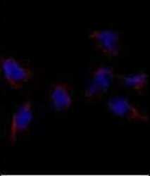
- Immunofluorescent analysis using a Bmp7 polyclonal antibody (Product # PA5-11720) in HeLa cells. 0.025 mg/mL primary antibody was followed by fluor-conjugated donkey anti-rabbit lgG (H+L) (orange). Blue counterstaining is DAPI.

- Submitted by
- Invitrogen Antibodies (provider)
- Main image

- Experimental details
- Immunofluorescence analysis of Bmp7 was performed using 70% confluent log phase MCF7 and HeLa cells. The cells were fixed with 4% paraformaldehyde for 10 minutes, permeabilized with 0.1% Triton™ X-100 for 15 minutes, and blocked with 2% BSA for 45 minutes at room temperature. The cells were labeled with BMP-7 Polyclonal Antibody (Product # PA5-11720) at 1:100 dilution in 0.1% BSA, incubated at 4 degree celsius overnight and then labeled with Donkey anti-Rabbit IgG (H+L) Highly Cross-Adsorbed Secondary Antibody, Alexa Fluor Plus 488 (Product # A32790, 1:2000 dilution), for 45 minutes at room temperature (Panel a: Green). Nuclei (Panel b: Blue) were stained with ProLong™ Diamond Antifade Mountant with DAPI (Product # P36962). F-actin (Panel c: Red) was stained with Rhodamine Phalloidin (Product # R415, 1:300). Panel d represents the merged image showing nuclear and cytoplasmic localization of BMP7 in MCF7. Panel e represents HeLa cells with lower expression of BMP7. Panel f represents control cells with no primary antibody to assess background. The images were captured at 60X magnification.

Supportive validation
- Submitted by
- Invitrogen Antibodies (provider)
- Main image

- Experimental details
- Immunohistochemical analysis of formalin-fixed, paraffin-embedded human cancer tissue using a Bmp7 polyclonal antibody (Product # PA5-11720), followed by HRP-conjugated secondary antibody and DAB staining.
