 Explore
 Validate
 Learn
Learn Western blot
Antibody data
- Antibody Data
- Antigen structure
- References [1]
- Comments [0]
- Validations
- Western blot [1]
- Immunohistochemistry [2]
- Other assay [1]
Submit
Validation data
Reference
Comment
Report error
- Product number
- PA5-79722 - Provider product page

- Provider
- Invitrogen Antibodies
- Product name
- NDRG2 Polyclonal Antibody
- Antibody type
- Polyclonal
- Antigen
- Synthetic peptide
- Description
- Reconstitute with 0.2 mL of distilled water to yield a concentration of 500 µg/mL.
- Reactivity
- Human, Mouse, Rat
- Host
- Rabbit
- Isotype
- IgG
- Vial size
- 100 µg
- Concentration
- 500 µg/mL
- Storage
- -20°C
Submitted references Cell type-specific biotin labeling in vivo resolves regional neuronal and astrocyte proteomic differences in mouse brain.
Rayaprolu S, Bitarafan S, Santiago JV, Betarbet R, Sunna S, Cheng L, Xiao H, Nelson RS, Kumar P, Bagchi P, Duong DM, Goettemoeller AM, Oláh VJ, Rowan M, Levey AI, Wood LB, Seyfried NT, Rangaraju S
Nature communications 2022 May 25;13(1):2927
Nature communications 2022 May 25;13(1):2927
No comments: Submit comment
Supportive validation
- Submitted by
- Invitrogen Antibodies (provider)
- Main image
- Experimental details
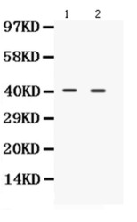
- Western blot analysis of NDRG2 in Lane 1: rat brain tissue lysate, Lane 2: mouse brain tissue lysate using 50 µg per well. Sample was incubated with NDRG2 (Product # PA5-79722) at a dilution of 0.5 µg/mL.

Supportive validation
- Submitted by
- Invitrogen Antibodies (provider)
- Main image
- Experimental details
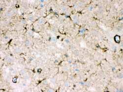
- Immunohistochemistry analysis of NDRG2 on paraffin-embedded mouse brain tissue. Sample was incubated with NDRG2 polyclonal antibody (Product# PA5-79722).
- Submitted by
- Invitrogen Antibodies (provider)
- Main image
- Experimental details
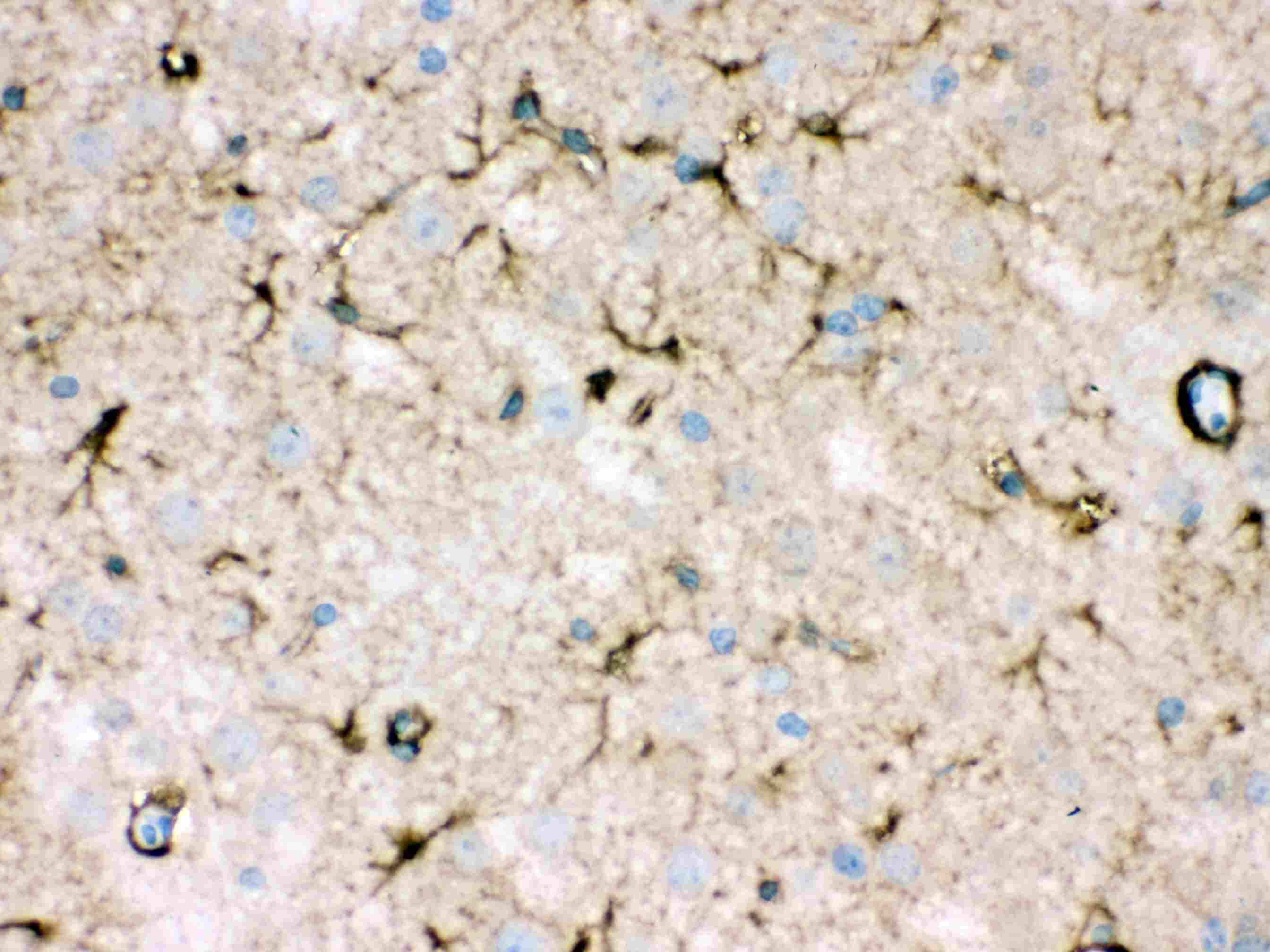
- Immunohistochemistry analysis of NDRG2 on paraffin-embedded rat brain tissue. Sample was incubated with NDRG2 polyclonal antibody (Product# PA5-79722).

Supportive validation
- Submitted by
- Invitrogen Antibodies (provider)
- Main image
- Experimental details
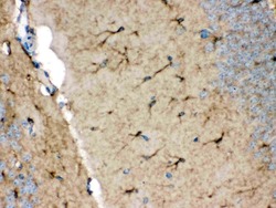
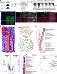
- Astrocyte protein biotinylation reveals region-specific proteomic signatures and differences between Camk2a neurons. a Study design for neuron-specific and astrocyte-specific proteomic biotinylation. Rosa26 TurboID/wt , Rosa26 TurboID/wt /Camk2a-Cre Ert2 , Rosa26 TurboID/wt /Aldh1l1-Cre Ert2 mice received tamoxifen intraperitoneally for 5 days. After 4 weeks, mice received biotin water for 2 weeks. b Representative Western blots from brain region lysates ( n = 2 mice/group), probed for biotin (streptavidin Alexa488), V5 and Gapdh are shown. c Representative images ( n = 2 mice/group) showing biotinylation in the hippocampus. d Representative immunofluorescence images ( n = 2 mice/group) showing overlap between astrocytic biotinylation (streptavidin Alexa488), Gfap, and Ndrg2 in the hippocampus region from Rosa26 TurboID/wt /Aldh1l1 mice. e Representative immunofluorescence images ( n = 2 mice/group) showing no overlap between biotinylation, Iba1, and betaIII-tubulin in astrocytes and blood vessels in the hippocampus region from Rosa26 TurboID/wt /Aldh1l1 mice. f Clustering analysis of protein abundance data of region-enriched proteins with at least 4-fold enrichment over other regions in Rosa26 TurboID/wt /Aldh1l1 mice. STRING analysis identified networks of direct (protein-protein) and indirect (functional) interactions within core regional protein signatures in cortex/hippocampus and cerebellum. g Heatmap representation, based on enrichment Z-scores, of gene ontologies enri
