 Explore
 Validate
 Learn
Learn Western blot
 Immunocytochemistry
Antibody data
- Antibody Data
- Antigen structure
- References [0]
- Comments [0]
- Validations
- Immunocytochemistry [3]
- Immunoprecipitation [1]
- Immunohistochemistry [1]
- Chromatin Immunoprecipitation [2]
- Other assay [1]
Submit
Validation data
Reference
Comment
Report error
- Product number
- PA5-29106 - Provider product page

- Provider
- Invitrogen Antibodies
- Product name
- MCM3 Polyclonal Antibody
- Antibody type
- Polyclonal
- Antigen
- Recombinant full-length protein
- Description
- Recommended positive controls: 293T, A431, H1299, HeLa, HepG2, Molt-4, Raji. Predicted reactivity: Mouse (95%), Rat (94%), Zebrafish (80%), Xenopus laevis (84%), Chicken (86%), Rhesus Monkey (99%), Bovine (97%). Store product as a concentrated solution. Centrifuge briefly prior to opening the vial.
- Reactivity
- Human, Mouse
- Host
- Rabbit
- Isotype
- IgG
- Vial size
- 100 μL
- Concentration
- 0.71 mg/mL
- Storage
- Store at 4°C short term. For long term storage, store at -20°C, avoiding freeze/thaw cycles.
No comments: Submit comment
Supportive validation
- Submitted by
- Invitrogen Antibodies (provider)
- Main image
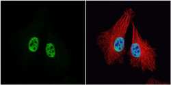
- Experimental details
- Immunocytochemistry-Immunofluorescence analysis of MCM3 was performed in HeLa cells fixed in 4% paraformaldehyde at RT for 15 min. Green: MCM3 Polyclonal Antibody (Product # PA5-29106) diluted at 1:500. Red: alpha Tubulin, a cytoskeleton marker. Blue: Hoechst 33342 staining.
- Submitted by
- Invitrogen Antibodies (provider)
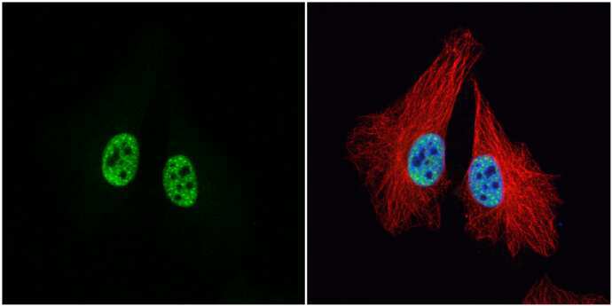
- Main image

- Experimental details
- Immunocytochemistry-Immunofluorescence analysis of MCM3 was performed in HeLa cells fixed in 4% paraformaldehyde at RT for 15 min. Green: MCM3 Polyclonal Antibody (Product # PA5-29106) diluted at 1:500. Red: alpha Tubulin, a cytoskeleton marker. Blue: Hoechst 33342 staining.

- Submitted by
- Invitrogen Antibodies (provider)
- Main image
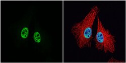
- Experimental details
- Immunocytochemistry-Immunofluorescence analysis of MCM3 was performed in HeLa cells fixed in 4% paraformaldehyde at RT for 15 min. Green: MCM3 Polyclonal Antibody (Product # PA5-29106) diluted at 1:500. Red: alpha Tubulin, a cytoskeleton marker. Blue: Hoechst 33342 staining.
Supportive validation
- Submitted by
- Invitrogen Antibodies (provider)
- Main image
- Experimental details
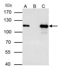
- MCM3 Polyclonal Antibody immunoprecipitates MCM3 protein in IP experiments. IP samples: HepG2 whole cell extract. A. 40 µg HepG2 whole cell extract. B. Control with 4 µg of preimmune Rabbit IgG. C. Immunoprecipitation of MCM3 protein by 4 µg MCM3 Polyclonal Antibody (Product # PA5-29106). 7.5 % SDS-PAGE. The immunoprecipitated MCM3 protein was detected by MCM3 Polyclonal Antibody (Product # PA5-29106) diluted at 1:1,000.

Supportive validation
- Submitted by
- Invitrogen Antibodies (provider)
- Main image

- Experimental details
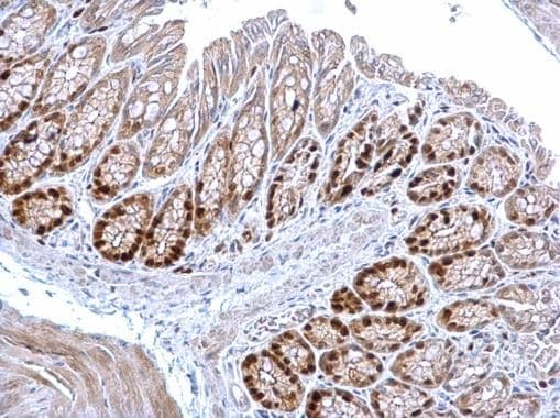
- MCM3 Polyclonal Antibody detects MCM3 protein at nucleus on mouse colon by immunohistochemical analysis. Sample: Paraffin-embedded mouse colon. MCM3 Polyclonal Antibody (Product # PA5-29106) diluted at 1:1,000. Antigen Retrieval: EDTA based buffer, pH 8.0, 15 min.
Supportive validation
- Submitted by
- Invitrogen Antibodies (provider)
- Main image

- Experimental details
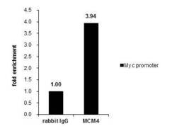
- Cross-linked ChIP was performed with HeLa chromatin extract and 5 µg of either control rabbit IgG or MCM3 Polyclonal Antibody (Product # PA5-29106). The precipitated DNA was detected by PCR with primer set targeting to Myc promoter.

- Submitted by
- Invitrogen Antibodies (provider)
- Main image
- Experimental details
- Cross-linked ChIP was performed with HeLa chromatin extract and 5 µg of either control rabbit IgG or MCM3 Polyclonal Antibody (Product # PA5-29106). The precipitated DNA was detected by PCR with primer set targeting to Myc promoter.

Supportive validation
- Submitted by
- Invitrogen Antibodies (provider)
- Main image

- Experimental details
- MCM3 Polyclonal Antibody immunoprecipitates MCM3 protein in IP experiments. IP samples: HepG2 whole cell extract. A. 40 µg HepG2 whole cell extract. B. Control with 4 µg of preimmune Rabbit IgG. C. Immunoprecipitation of MCM3 protein by 4 µg MCM3 Polyclonal Antibody (Product # PA5-29106). 7.5 % SDS-PAGE. The immunoprecipitated MCM3 protein was detected by MCM3 Polyclonal Antibody (Product # PA5-29106) diluted at 1:1,000.
