Explore
 Validate
 Learn
Learn Western blot
 Immunohistochemistry
Antibody data
- Antibody Data
- Antigen structure
- References [1]
- Comments [0]
- Validations
- Immunohistochemistry [1]
- Other assay [5]
Submit
Validation data
Reference
Comment
Report error
- Product number
- PA5-14421 - Provider product page

- Provider
- Invitrogen Antibodies
- Product name
- JNK3 Polyclonal Antibody
- Antibody type
- Polyclonal
- Antigen
- Synthetic peptide
- Reactivity
- Human, Mouse
- Host
- Rabbit
- Isotype
- IgG
- Vial size
- 400 μL
- Concentration
- 2 mg/mL
- Storage
- Store at 4°C short term. For long term storage, store at -20°C, avoiding freeze/thaw cycles.
Submitted references Colocalization and Interaction Study of Neuronal JNK3, JIP1, and β-Arrestin2 Together with PSD95.
Musi CA, Marchini G, Giani A, Tomaselli G, Priori EC, Colnaghi L, Borsello T
International journal of molecular sciences 2022 Apr 8;23(8)
International journal of molecular sciences 2022 Apr 8;23(8)
No comments: Submit comment
Supportive validation
- Submitted by
- Invitrogen Antibodies (provider)
- Main image
- Experimental details
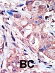
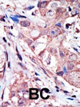
- Immunohistochemistry analysis of JNK3 in formalin-fixed and paraffin-embedded human cancer tissue. Samples were incubated with JNK3 polyclonal antibody (Product # PA5-14421) which was peroxidase-conjugated to the secondary antibody, followed by AEC staining. This data demonstrates the use of this antibody for immunohistochemistry; clinical relevance has not been evaluated. BC = breast carcinoma; HC = hepatocarcinoma.
Supportive validation
- Submitted by
- Invitrogen Antibodies (provider)
- Main image
- Experimental details
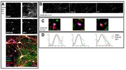
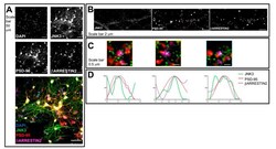
- Confocal microscopy and SIM outputs used to assess JNK3-JIP1-PSD95 colocalization. ( A ) Neurons were fixed and stained with anti-JNK3 (green), anti-PSD95 (red), and anti-JIP1 (violet). Images were obtained using a Nikon N-SIM confocal microscope and overlaid to assess protein localization. Nuclei were stained with Hoechst. Scale bar of 50 mum. ( B ) 3D-SIM images were acquired with a 100x objective. Scale bar of 2 mum. Squares represent the location of the inset present in panel ( B ). ( C ) Merged images of reconstructed 3D-SIM images of JNK3 (green), PSD95 (red), and JIP1 (violet). Scale bar of 0.5 mum. ( D ) Intensity profile (green for JNK3, red for PSD95, violet for JIP1) representing the values indicated by the arrows in panel ( C ). The values are normalized to 100 (arbitrary unit).

- Submitted by
- Invitrogen Antibodies (provider)
- Main image
- Experimental details
- Confocal microscopy and SIM outputs used to assess JNK3-beta-arrestin2-PSD95 colocalization. ( A ) Neurons were fixed and stained with anti-JNK3 (green), anti-PSD95 (red), and anti-beta-arrestin2 (violet). Images were obtained using a Nikon N-SIM confocal microscope and overlaid to assess protein localization. Nuclei were stained with Hoechst. Scale bar of 50 mum. ( B ) 3D-SIM images were acquired with a 100x objective. Scale bar of 2 mum. Squares represent the location of the inset present in panel ( B ). ( C ) Merged images of reconstructed 3D-SIM images of JNK3 (green), PSD95 (red), and beta-arrestin2 (violet). Scale bar of 0.5 mum. ( D ) The intensity profile (green for JNK3, red for PSD95, violet for beta-arrestin2) represents the values indicated by the arrows in panel ( C ). The values are normalized to 100 (arbitrary unit).

- Submitted by
- Invitrogen Antibodies (provider)
- Main image
- Experimental details
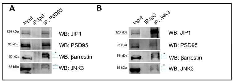
- Immunoprecipitation outputs used to assess JNK3-JIP1-beta-arrestin2-PSD95 colocalization in primary hippocampal neurons. ( A ) PSD95 was immunoprecipitated from the cell lysate homogenate using PSD95 antibody, and immune complexes were analyzed for the presence of JNK3, JIP1, and beta-arrestin2. Immunoprecipitation with IgG antibody was used as a control. ( B ) JNK3 was immunoprecipitated from the cell lysate homogenate using JNK3-specific antibody, separated by SDS-PAGE, and analyzed via Western blot with anti-PSD95, anti-JIP1, and anti-beta-arrestin2 antibodies. Immunoprecipitation with IgG antibody was used as a control. * indicates non-specific bands.
- Submitted by
- Invitrogen Antibodies (provider)
- Main image
- Experimental details
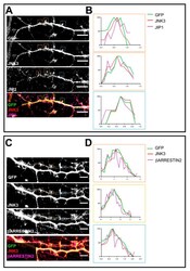
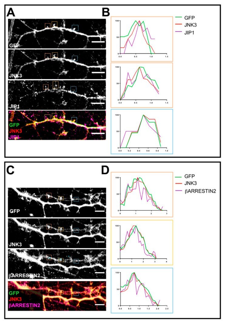
- Confocal microscopy outputs used to assess JNK3-JIP1-beta-arrestin2-PSD95 colocalization at the dendritic spine level. ( A ) Neurons were fixed and stained with anti-JNK3 (red), anti-JIP1 (violet), and anti-GFP (green). Images were obtained using a Nikon N-SIM confocal microscope and overlaid to assess protein localization. Scale bar of 50 mum. Squares represent the loci analyzed in panel ( B ). ( B ) Intensity profile (green for GFP, red for JNK3, violet for JIP1) representing the values indicated by the squares in panel ( A ). The values are normalized to 100 (arbitrary unit). ( C ) Neurons were fixed and stained with anti-JNK3 (red), anti-beta-arrestin2 (violet), and anti-GFP (green). Images were obtained using a Nikon N-SIM confocal microscope and overlaid to assess protein localization. Scale bar of 50 mum. Squares represent the loci analyzed in panel ( B ). ( D ) Intensity profile (green for GFP, red for JNK3, violet for beta-arrestin2) representing the values indicated by the squares in panel ( A ). The values are normalized to 100 (arbitrary unit).
- Submitted by
- Invitrogen Antibodies (provider)
- Main image
- Experimental details
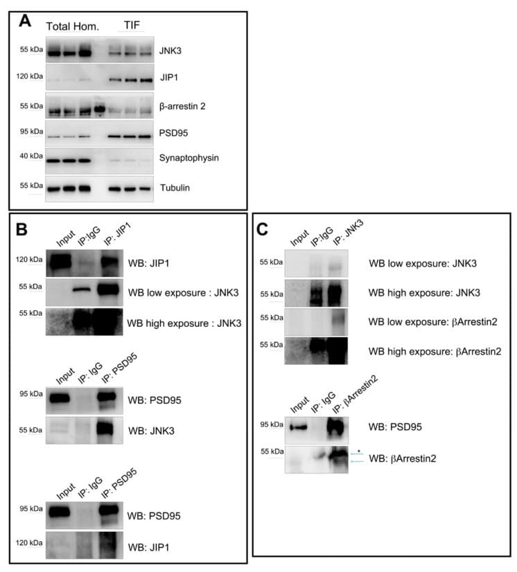
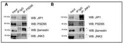
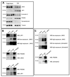
- Immunoprecipitation outputs used to assess JNK3-JIP1-beta-arrestin2-PSD95 interactions at the synapse. ( A ) Western blots performed on total brain homogenate and Triton-insoluble fraction (TIF) reveal the different amounts of JNK3, JIP1, beta-arrestin2, and PSD95 in the two extracts analyzed. Synaptophysin was used as a control for the purity of the TIF preparation. ( B ) JIP1 was immunoprecipitated from the TIF using JIP1 antibody, and immune complexes were analyzed for the presence of JNK3 (rabbit). Immunoprecipitation with IgG antibody was used as a control. PSD95 was immunoprecipitated from the TIF using PSD95-specific antibody, separated by SDS-PAGE, and analyzed with Western blot with anti-PSD95, anti-JNK3 (mouse), and anti-JIP1 antibodies. Immunoprecipitation with IgG antibody was used as a control. ( C ) JNK3 was immunoprecipitated from the TIF using the JNK3 antibody, and immune complexes were analyzed for the presence of beta-arrestin2 and JNK3 (rabbit). Immunoprecipitation with IgG antibody was used as a control. beta-arrestin2 was immunoprecipitated from the TIF using beta-arrestin2-specific antibody, separated by SDS-PAGE, and analyzed by Western blot with anti-PSD95, anti-JNK3, and anti-beta-arrestin2 antibodies. Immunoprecipitation with IgG antibody was used as a control. * indicates non-specific bands.