Explore
 Validate
 Learn
LearnMA1-23269
antibody from Invitrogen Antibodies
Targeting: RAD50
hRad50, RAD50-2
 Western blot
 Immunocytochemistry
 Immunoprecipitation Immunohistochemistry Chromatin Immunoprecipitation Other assay
Antibody data
- Antibody Data
- Antigen structure
- References [2]
- Comments [0]
- Validations
- Immunocytochemistry [2]
- Immunohistochemistry [5]
- Other assay [1]
Submit
Validation data
Reference
Comment
Report error
- Product number
- MA1-23269 - Provider product page

- Provider
- Invitrogen Antibodies
- Product name
- RAD50 Monoclonal Antibody (13B3)
- Antibody type
- Monoclonal
- Antigen
- Other
- Description
- Recommended positive controls: HeLa, HeLa nuclear extract.
- Reactivity
- Human, Mouse, Rat
- Host
- Mouse
- Isotype
- IgG
- Antibody clone number
- 13B3
- Vial size
- 100 μL
- Concentration
- 1.7 mg/mL
- Storage
- Store at 4°C short term. For long term storage, store at -20°C, avoiding freeze/thaw cycles.
Submitted references Loss of tumor suppressor inositol polyphosphate 4-phosphatase type B impairs DNA double-strand break repair by destabilization of DNA tethering protein Rad50.
Positional stability of single double-strand breaks in mammalian cells.
Sun Y, Ning X, Fan J, Hu J, Jiang Y, Hu Z, Paulo JA, Liu J, Qiu X, Xu H, Fu S, Gygi SP, Zhang J, Zhou C
Cell death & disease 2020 Apr 27;11(4):292
Cell death & disease 2020 Apr 27;11(4):292
Positional stability of single double-strand breaks in mammalian cells.
Soutoglou E, Dorn JF, Sengupta K, Jasin M, Nussenzweig A, Ried T, Danuser G, Misteli T
Nature cell biology 2007 Jun;9(6):675-82
Nature cell biology 2007 Jun;9(6):675-82
No comments: Submit comment
Supportive validation
- Submitted by
- Invitrogen Antibodies (provider)
- Main image
- Experimental details
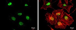
- Immunocytochemistry-Immunofluorescence analysis of RAD50 was performed in HeLa cells fixed in 4% paraformaldehyde at RT for 15 min. Green: RAD50 Monoclonal Antibody (13B3) (Product # MA1-23269) diluted at 1:200. Red: phalloidin, a cytoskeleton marker. Scale bar = 10 µm.
- Submitted by
- Invitrogen Antibodies (provider)
- Main image

- Experimental details
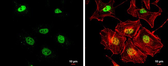
- Immunocytochemistry-Immunofluorescence analysis of RAD50 was performed in HeLa cells fixed in 4% paraformaldehyde at RT for 15 min. Green: RAD50 Monoclonal Antibody (13B3) (Product # MA1-23269) diluted at 1:200. Red: phalloidin, a cytoskeleton marker. Scale bar = 10 µm.

Supportive validation
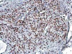
- Submitted by
- Invitrogen Antibodies (provider)
- Main image

- Experimental details
- Immunohistochemistry (Paraffin) analysis of RAD50 was performed in paraffin-embedded PC-3 xenograft tissue using RAD50 Monoclonal Antibody (13B3) (Product # MA1-23269) at a dilution of 1:200.
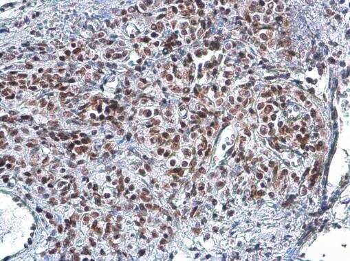
- Submitted by
- Invitrogen Antibodies (provider)
- Main image

- Experimental details
- Immunohistochemistry (Paraffin) analysis of RAD50 was performed in paraffin-embedded CAL 27 xenograft tissue using RAD50 Monoclonal Antibody (13B3) (Product # MA1-23269) at a dilution of 1:200.
- Submitted by
- Invitrogen Antibodies (provider)
- Main image

- Experimental details
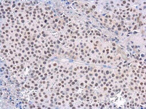
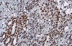
- Immunohistochemistry (Paraffin) analysis of RAD50 was performed in paraffin-embedded human lung tissue using RAD50 Monoclonal Antibody (13B3) (Product # MA1-23269) at a dilution of 1:200.
- Submitted by
- Invitrogen Antibodies (provider)
- Main image
- Experimental details
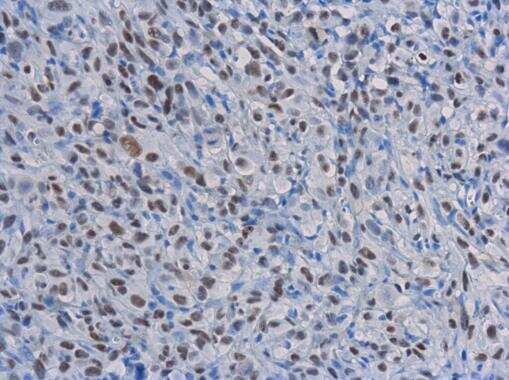
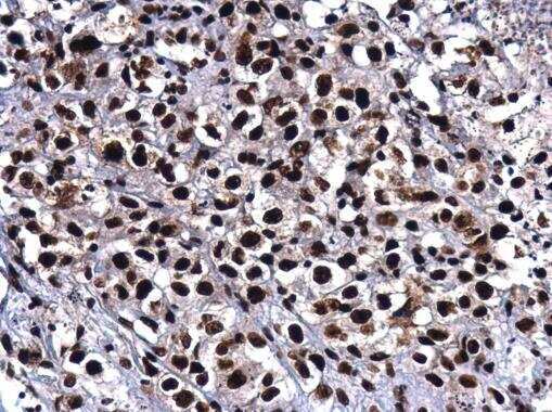
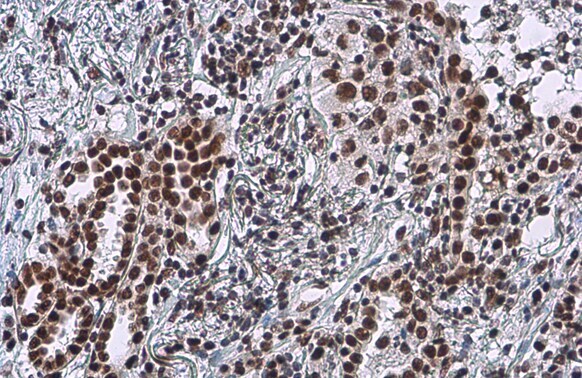
- Immunohistochemistry (Paraffin) analysis of RAD50 was performed in paraffin-embedded human lung cancer tissue using RAD50 Monoclonal Antibody (13B3) (Product # MA1-23269) at a dilution of 1:100. Antigen Retrieval: Citrate buffer, pH 6.0, 15 min.
- Submitted by
- Invitrogen Antibodies (provider)
- Main image
- Experimental details
- Immunohistochemistry (Paraffin) analysis of RAD50 was performed in paraffin-embedded human lung cancer tissue using RAD50 Monoclonal Antibody (13B3) (Product # MA1-23269) at a dilution of 1:100. Antigen Retrieval: Citrate buffer, pH 6.0, 15 min.
Supportive validation
- Submitted by
- Invitrogen Antibodies (provider)
- Main image

- Experimental details
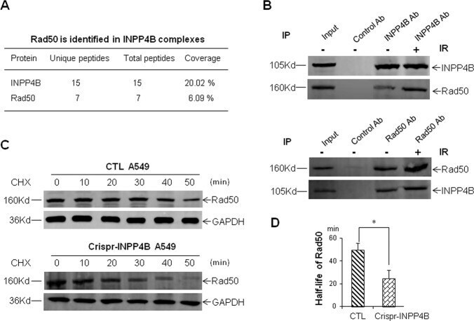
- Fig. 6 Interaction of INPP4B with Rad50 and destabilization of Rad50 upon loss of INPP4B. a Rad50 was identified from immuno-precipitated INPP4B complexes by mass spectrometry. b Reciprocal verification of the interaction between INPP4B and Rad50 in A549 cells. Two micrograms of antibodies against INPP4B or Rad50 were used for each reciprocal immunoprecipitation in a total of 2 mg of A549 cell lysates, and the precipitated proteins were resolved on 6% SDS-PAGE gel and detected with the indicated antibodies. In IR treatments, cells were harvested at 2 h post IR. c Representative Western blot images for measuring the stability of Rad50 in CTL and Crispr-INPP4B A549 cells. The half-life of Rad50 in each cell line was determined by densitometry scanning compared to the signal intensity at time zero with CHX treatment, and normalized to GAPDH signal. The average half-life of Rad50 was based on three independent CHX treatments and plotted in d , and data are presented as means +- SD (min). An error bar represents SD. Asterisk (*) indicates p < 0.05 by Student's t -test.