Explore
 Validate
 Learn
Learn Immunohistochemistry
Antibody data
- Antibody Data
- Antigen structure
- References [12]
- Comments [0]
- Validations
- Immunohistochemistry [4]
- Flow cytometry [6]
- Other assay [7]
Submit
Validation data
Reference
Comment
Report error
- Product number
- MA5-12345 - Provider product page

- Provider
- Invitrogen Antibodies
- Product name
- MUC2 Monoclonal Antibody (996/1)
- Antibody type
- Monoclonal
- Antigen
- Synthetic peptide
- Description
- MA5-12345 targets Mucin 2 in IHC (P), ICC/IF and FACS applications and shows reactivity with human and mouse samples. The MA5-12345 immunogen is mUC2 tandem repeat peptide.
- Reactivity
- Human, Mouse
- Host
- Mouse
- Isotype
- IgG
- Antibody clone number
- 996/1
- Vial size
- 500 μL
- Concentration
- 0.1 mg/mL
- Storage
- 4°C
Submitted references A Novel In Vitro Membrane Permeability Methodology Using Three-dimensional Caco-2 Tubules in a Microphysiological System Which Better Mimics In Vivo Physiological Conditions.
Human intestinal bitter taste receptors regulate innate immune responses and metabolic regulators in obesity.
Metformin Exerts Anti-inflammatory and Mucus Barrier Protective Effects by Enriching Akkermansia muciniphila in Mice With Ulcerative Colitis.
Parechovirus A Infection of the Intestinal Epithelium: Differences Between Genotypes A1 and A3.
Small proline-rich protein 2A is a gut bactericidal protein deployed during helminth infection.
Hyperglycemia minimally alters primary self-renewing human colonic epithelial cells while TNFα-promotes severe intestinal epithelial dysfunction.
Evaluation of a Live Attenuated S. sonnei Vaccine Strain in the Human Enteroid Model.
Host Specificity of Flagellins from Segmented Filamentous Bacteria Affects Their Patterns of Interaction with Mouse Ileal Mucosal Proteins.
Early Gastric Cancer-Like Advanced Gastric Cancer versus Advanced Gastric Cancer-Like Early Gastric Cancer.
Change of mucosal color in early gastric cancer.
MUC5AC, cytokeratin 20 and HER2 expression and K-RAS mutations within mucinogenic growth in congenital pulmonary airway malformations.
Unclassified mucin phenotype of gastric adenocarcinoma exhibits the highest invasiveness.
Hagiwara Y, Kumagai H, Ouwerkerk N, Gijzen L, Annida R, Bokkers M, van Vught R, Yoshinari K, Katakawa Y, Motonaga K, Tajiri T
Journal of pharmaceutical sciences 2022 Jan;111(1):214-224
Journal of pharmaceutical sciences 2022 Jan;111(1):214-224
Human intestinal bitter taste receptors regulate innate immune responses and metabolic regulators in obesity.
Liszt KI, Wang Q, Farhadipour M, Segers A, Thijs T, Nys L, Deleus E, Van der Schueren B, Gerner C, Neuditschko B, Ceulemans LJ, Lannoo M, Tack J, Depoortere I
The Journal of clinical investigation 2022 Feb 1;132(3)
The Journal of clinical investigation 2022 Feb 1;132(3)
Metformin Exerts Anti-inflammatory and Mucus Barrier Protective Effects by Enriching Akkermansia muciniphila in Mice With Ulcerative Colitis.
Ke H, Li F, Deng W, Li Z, Wang S, Lv P, Chen Y
Frontiers in pharmacology 2021;12:726707
Frontiers in pharmacology 2021;12:726707
Parechovirus A Infection of the Intestinal Epithelium: Differences Between Genotypes A1 and A3.
García-Rodríguez I, van Eijk H, Koen G, Pajkrt D, Sridhar A, Wolthers KC
Frontiers in cellular and infection microbiology 2021;11:740662
Frontiers in cellular and infection microbiology 2021;11:740662
Small proline-rich protein 2A is a gut bactericidal protein deployed during helminth infection.
Hu Z, Zhang C, Sifuentes-Dominguez L, Zarek CM, Propheter DC, Kuang Z, Wang Y, Pendse M, Ruhn KA, Hassell B, Behrendt CL, Zhang B, Raj P, Harris-Tryon TA, Reese TA, Hooper LV
Science (New York, N.Y.) 2021 Nov 5;374(6568):eabe6723
Science (New York, N.Y.) 2021 Nov 5;374(6568):eabe6723
Hyperglycemia minimally alters primary self-renewing human colonic epithelial cells while TNFα-promotes severe intestinal epithelial dysfunction.
Dutton JS, Hinman SS, Kim R, Attayek PJ, Maurer M, Sims CS, Allbritton NL
Integrative biology : quantitative biosciences from nano to macro 2021 Jun 15;13(6):139-152
Integrative biology : quantitative biosciences from nano to macro 2021 Jun 15;13(6):139-152
Evaluation of a Live Attenuated S. sonnei Vaccine Strain in the Human Enteroid Model.
Pilla G, Wu T, Grassel C, Moon J, Foulke-Abel J, Tang CM, Barry EM
Pathogens (Basel, Switzerland) 2021 Aug 25;10(9)
Pathogens (Basel, Switzerland) 2021 Aug 25;10(9)
Host Specificity of Flagellins from Segmented Filamentous Bacteria Affects Their Patterns of Interaction with Mouse Ileal Mucosal Proteins.
Chen H, Yin Y, Wang Y, Wang X, Xiang C
Applied and environmental microbiology 2017 Sep 15;83(18)
Applied and environmental microbiology 2017 Sep 15;83(18)
Early Gastric Cancer-Like Advanced Gastric Cancer versus Advanced Gastric Cancer-Like Early Gastric Cancer.
Park HS, Lee SY, Hong SN, Kim JH, Sung IK, Park HS, Shim CS, Jin CJ
Clinical endoscopy 2013 Mar;46(2):155-60
Clinical endoscopy 2013 Mar;46(2):155-60
Change of mucosal color in early gastric cancer.
Lee HH, Lee SY, Yoon HY, Hong SN, Kim JH, Sung IK, Park HS, Shim CS, Han HS
Journal of digestive diseases 2012 Oct;13(10):510-6
Journal of digestive diseases 2012 Oct;13(10):510-6
MUC5AC, cytokeratin 20 and HER2 expression and K-RAS mutations within mucinogenic growth in congenital pulmonary airway malformations.
Rossi G, Gasser B, Sartori G, Migaldi M, Costantini M, Mengoli MC, Piccioli S, Cavazza A, Rivasi F
Histopathology 2012 Jun;60(7):1133-43
Histopathology 2012 Jun;60(7):1133-43
Unclassified mucin phenotype of gastric adenocarcinoma exhibits the highest invasiveness.
Han HS, Lee SY, Lee KY, Hong SN, Kim JH, Sung IK, Park HS, Jin CJ, Min YI
Journal of gastroenterology and hepatology 2009 Apr;24(4):658-66
Journal of gastroenterology and hepatology 2009 Apr;24(4):658-66
No comments: Submit comment
Supportive validation
- Submitted by
- Invitrogen Antibodies (provider)
- Main image
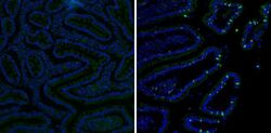
- Experimental details
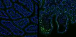
- Immunofluorescent analysis of Mucin 2 (green) showing staining in the cytoplasm of human small intestine tissue (right) compared to a negative control without primary antibody (left). Formalin-fixed tissue was permeabilized with 0.1% Triton X-100 in TBS for 5-10 minutes and blocked with 3% BSA-PBS for 30 minutes at room temperature. Tissue was probed with a Mucin 2 monoclonal antibody (Product # MA5-12345) in 3% BSA-PBS at a dilution of 1:100 and incubated overnight at 4ºC in a humidified chamber. Tissue was washed with PBST and incubated with a DyLight-conjugated secondary antibody in PBS at room temperature in the dark. Nuclei were stained with Hoechst or DAPI (blue). Images were taken at a magnification of 60x.
- Submitted by
- Invitrogen Antibodies (provider)
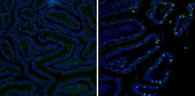
- Main image

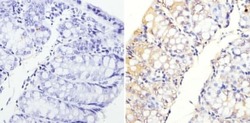
- Experimental details
- Immunohistochemistry analysis of Mucin 2 showing staining in the cytoplasm of paraffin-embedded human colon tissue (right) compared to a negative control without primary antibody (left). To expose target proteins, antigen retrieval was performed using 10mM sodium citrate (pH 6.0), microwaved for 8-15 min. Following antigen retrieval, tissues were blocked in 3% H2O2-methanol for 15 min at room temperature, washed with ddH2O and PBS, and then probed with a Mucin 2 monoclonal antibody (Product # MA5-12345) diluted in 3% BSA-PBS at a dilution of 1:200 overnight at 4°C in a humidified chamber. Tissues were washed extensively in PBST and detection was performed using an HRP-conjugated secondary antibody followed by colorimetric detection using a DAB kit. Tissues were counterstained with hematoxylin and dehydrated with ethanol and xylene to prep for mounting.
- Submitted by
- Invitrogen Antibodies (provider)
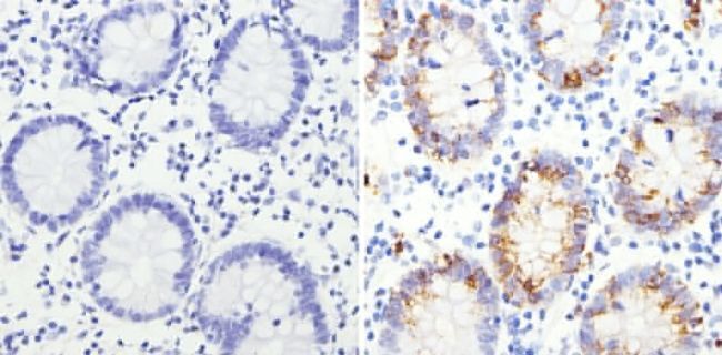
- Main image
- Experimental details
- Immunohistochemistry analysis of Mucin 2 showing staining in the cytoplasm of paraffin-embedded mouse colon tissue (right) compared to a negative control without primary antibody (left). To expose target proteins, antigen retrieval was performed using 10mM sodium citrate (pH 6.0), microwaved for 8-15 min. Following antigen retrieval, tissues were blocked in 3% H2O2-methanol for 15 min at room temperature, washed with ddH2O and PBS, and then probed with a Mucin 2 monoclonal antibody (Product # MA5-12345) diluted in 3% BSA-PBS at a dilution of 1:20 overnight at 4°C in a humidified chamber. Tissues were washed extensively in PBST and detection was performed using an HRP-conjugated secondary antibody followed by colorimetric detection using a DAB kit. Tissues were counterstained with hematoxylin and dehydrated with ethanol and xylene to prep for mounting.

- Submitted by
- Invitrogen Antibodies (provider)
- Main image
- Experimental details
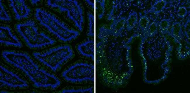
- Immunofluorescent analysis of Mucin 2 (green) showing staining in the cytoplasm of human colon tissue (right) compared to a negative control without primary antibody (left). Formalin-fixed tissue was permeabilized with 0.1% Triton X-100 in TBS for 5-10 minutes and blocked with 3% BSA-PBS for 30 minutes at room temperature. Tissue was probed with a Mucin 2 monoclonal antibody (Product # MA5-12345) in 3% BSA-PBS at a dilution of 1:100 and incubated overnight at 4ºC in a humidified chamber. Tissue was washed with PBST and incubated with a DyLight-conjugated secondary antibody in PBS at room temperature in the dark. Nuclei were stained with Hoechst or DAPI (blue). Images were taken at a magnification of 60x.
Supportive validation
- Submitted by
- Invitrogen Antibodies (provider)
- Main image
- Experimental details
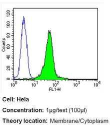
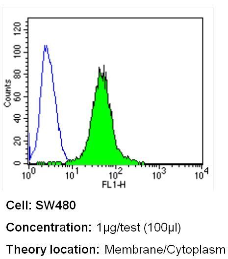
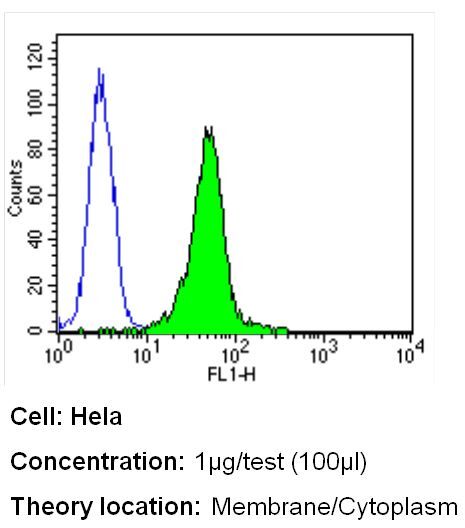
- Flow cytometry analysis of Mucin 2 in Hela cells (green) compared to an isotype control (blue). Cells were harvested, adjusted to a concentration of 1-5x10^6 cells/mL, fixed with 2% paraformaldehyde and washed with PBS. Cells were blocked with a 2% solution of BSA-PBS for 30 min at room temperature and incubated with a Mucin 2 monoclonal antibody (Product # MA5-12345) at a dilution of 1 µg/test for 40 min at room temperature. Cells were then incubated for 40 min at room temperature in the dark using a Dylight 488-conjugated secondary antibody and re-suspended in PBS for FACS analysis.

- Submitted by
- Invitrogen Antibodies (provider)
- Main image
- Experimental details
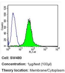
- Flow cytometry analysis of Mucin 2 in SW480 cells (green) compared to an isotype control (blue). Cells were harvested, adjusted to a concentration of 1-5x10^6 cells/mL, fixed with 2% paraformaldehyde and washed with PBS. Cells were blocked with a 2% solution of BSA-PBS for 30 min at room temperature and incubated with a Mucin 2 monoclonal antibody (Product # MA5-12345) at a dilution of 1 µg/test for 40 min at room temperature. Cells were then incubated for 40 min at room temperature in the dark using a Dylight 488-conjugated secondary antibody and re-suspended in PBS for FACS analysis.

- Submitted by
- Invitrogen Antibodies (provider)
- Main image
- Experimental details
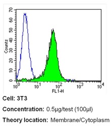
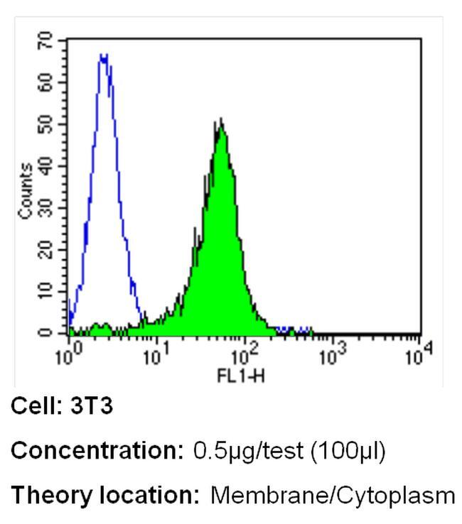
- Flow cytometry analysis of Mucin 2 in NIH-3T3 cells (green) compared to an isotype control (blue). Cells were harvested, adjusted to a concentration of 1-5x10^6 cells/mL, fixed with 2% paraformaldehyde and washed with PBS. Cells were blocked with a 2% solution of BSA-PBS for 30 min at room temperature and incubated with a Mucin 2 monoclonal antibody (Product # MA5-12345) at a dilution of 0.5 µg/test for 40 min at room temperature. Cells were then incubated for 40 min at room temperature in the dark using a Dylight 488-conjugated secondary antibody and re-suspended in PBS for FACS analysis.

- Submitted by
- Invitrogen Antibodies (provider)
- Main image
- Experimental details
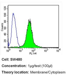
- Flow cytometry analysis of Mucin 2 in SW480 cells (green) compared to an isotype control (blue). Cells were harvested, adjusted to a concentration of 1-5x10^6 cells/mL, fixed with 2% paraformaldehyde and washed with PBS. Cells were blocked with a 2% solution of BSA-PBS for 30 min at room temperature and incubated with a Mucin 2 monoclonal antibody (Product # MA5-12345) at a dilution of 1 µg/test for 40 min at room temperature. Cells were then incubated for 40 min at room temperature in the dark using a Dylight 488-conjugated secondary antibody and re-suspended in PBS for FACS analysis.
- Submitted by
- Invitrogen Antibodies (provider)
- Main image

- Experimental details
- Flow cytometry analysis of Mucin 2 in NIH-3T3 cells (green) compared to an isotype control (blue). Cells were harvested, adjusted to a concentration of 1-5x10^6 cells/mL, fixed with 2% paraformaldehyde and washed with PBS. Cells were blocked with a 2% solution of BSA-PBS for 30 min at room temperature and incubated with a Mucin 2 monoclonal antibody (Product # MA5-12345) at a dilution of 0.5 µg/test for 40 min at room temperature. Cells were then incubated for 40 min at room temperature in the dark using a Dylight 488-conjugated secondary antibody and re-suspended in PBS for FACS analysis.
- Submitted by
- Invitrogen Antibodies (provider)
- Main image

- Experimental details
- Flow cytometry analysis of Mucin 2 in Hela cells (green) compared to an isotype control (blue). Cells were harvested, adjusted to a concentration of 1-5x10^6 cells/mL, fixed with 2% paraformaldehyde and washed with PBS. Cells were blocked with a 2% solution of BSA-PBS for 30 min at room temperature and incubated with a Mucin 2 monoclonal antibody (Product # MA5-12345) at a dilution of 1 µg/test for 40 min at room temperature. Cells were then incubated for 40 min at room temperature in the dark using a Dylight 488-conjugated secondary antibody and re-suspended in PBS for FACS analysis.
Supportive validation
- Submitted by
- Invitrogen Antibodies (provider)
- Main image

- Experimental details
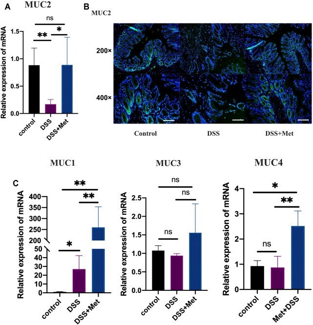
- FIGURE 2 Metformin protects the mucus barrier by increasing the expression of mucins in ulcerative colitis (UC). RT-qPCR assay of mucin 2 (MUC2) expression of colon tissue ( n = 3-4) (A) , immunofluorescence assay showing the expression of mucin2 protein in colon tissue (scale bars, 200 mum) ( n = 4) (B) , and RT-qPCR assay ( n = 3-6) of the relative expression levels of MUC1, MUC3, and MUC4 in colon tissue at mRNA level (C) and statistical comparison was made using an ANOVA test followed by Tukey-Kramer post-hoc tests. * p < 0.05, ** p < 0.01, *** p < 0.001, ns, no significance.
- Submitted by
- Invitrogen Antibodies (provider)
- Main image
- Experimental details
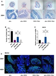
- FIGURE 6 Antibiotics abolish the mucus-protective effects of metformin. Alcian blue-periodic acid-Shiff (AB-PAS) staining (scale bars, 400 mum) ( n = 5-7) (A) and count of goblet cells ( n = 5-7) (B) , relative expression levels of mucin 2 (MUC2) ( n = 3-4) (C) , and immunofluorescence assay of mucin2 protein in the mouse colon (scale bars, 200 mum) ( n = 3) (D) and statistical comparison was made using an ANOVA test followed by Dunnett's post-hoc tests. * p < 0.05, ** p < 0.01, *** p < 0.001.

- Submitted by
- Invitrogen Antibodies (provider)
- Main image
- Experimental details
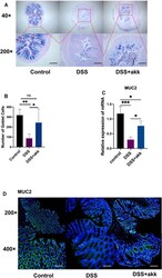
- FIGURE 8 Akkermansia muciniphila protects the mucus barrier in ulcerative colitis (UC). Alcian blue-periodic acid-Shiff (AB-PAS) staining of colon tissue (scale bars, 400 mum) ( n = 6-7) (A) and count of goblet cells ( n = 6-7) (B) , RT-qPCR assay of mucin 2 (MUC2) ( n = 3-4) (C) , and immunofluorescence assay of mucin2 protein detection in colon tissue (scale bars, 200 mum) ( n = 4) (D) and statistical comparison was made using an ANOVA test followed by Tukey-Kramer post-hoc tests. * p < 0.05, ** p < 0.01, *** p < 0.001, ns, no significance.
- Submitted by
- Invitrogen Antibodies (provider)
- Main image
- Experimental details
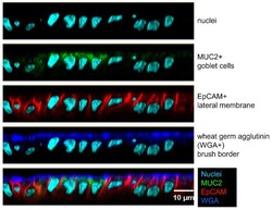
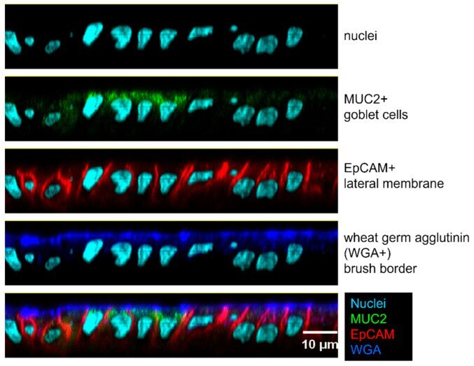
- Figure 2 Human enteroid monolayers include multiple cell types. The enteroid monolayer was fixed with methanol and stained with Hoechst for nuclei (light blue), antibodies against MUC2 for goblet cells (green), epithelial cell adhesion molecule (EpCAM) for lateral membranes (red), and wheat germ agglutinin (WGA) for brush border (blue).
- Submitted by
- Invitrogen Antibodies (provider)
- Main image
- Experimental details
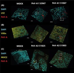
- Figure 6 Confocal images of infected cultures with clinical isolates of PeV-A1 and PeV-A3. In panel (A) , cultures were stained with Paneth cell marker (LYZ) in yellow and PeV-A antibody in red; in panel (B) , they were stained with enterocyte marker (villin) in yellow and PeV-A antibody in red, and in panel (C) , cultures were stained with goblet cell marker (MUC2) in yellow and PeV-A antibody in red. In all cases, nuclei were stained with DAPI in cyan. Scale bars in white represent 20 um, and the boxes indicate positively stained cells for the cell marker in the MOCK cases and for viral and cell marker staining in the infected images.
- Submitted by
- Invitrogen Antibodies (provider)
- Main image
- Experimental details
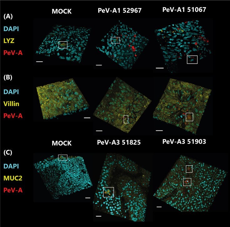
- Figure 1 Colocalization of TAS2R43 and TAS2R10 with Paneth or goblet cells in primary crypts from patients with obesity. Representative double-immunofluorescence images of jejunal crypts from obese patients. Crypts were stained for TAS2R43 ( A and B ) or TAS2R10 ( C and D ) (red) and alpha-defensin 5 (in Paneth cells) or mucin 2 (in goblet cells) (green). Images in A - C show colocalization, whereas the images in D show a TAS2R10 + cell in close proximity to a goblet cell, but no colocalization. Nuclei were stained with DAPI (blue). Scale bars: 20 mum ( C ) and 50 mum ( A , B , and D ). Each colocalization study was repeated in crypts derived from at least 3 patients with obesity.
- Submitted by
- Invitrogen Antibodies (provider)
- Main image

- Experimental details
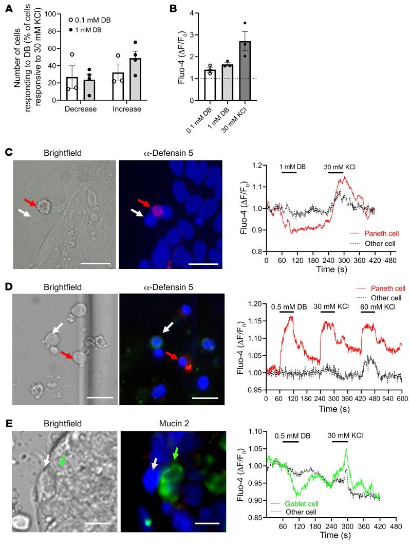
- Intracellular Ca 2+ changes in response to DB and immunostaining identification of cells in primary crypts from patients with obesity. ( A ) Percentage of 30 mM KCl-responsive primary jejunal cells from patients with obesity that responded to 0.1 mM or 1 mM DB administration with either a decrease or increase in intracellular Ca 2+ levels. ( B ) Relative rise in fluorescence intensity in single cells from crypts of individuals with obesity treated with DB or KCl. Data represent the mean +- SEM. n = 3-4 subjects. ( C - E ) Images show the cells in bright-field, as well as immunofluorescence staining for alpha-defensin 5 (red) and mucin 2 (green). Nuclei were stained with DAPI (blue). Ca 2+ changes in the regions of interest from the images in C - E are shown in the tracings. ( C ) Tracing of a Paneth cell responding to 1 mM DB with a Ca 2+ rise and another unidentified, nonresponding cell. ( D ) Tracing of a Paneth cell responding to 0.5 mM DB with a Ca 2+ increase and another unidentified, nonresponding cell. ( E ) Tracing of a goblet cell responding to 0.5 mM DB with a Ca 2+ decrease and an unidentified, nonresponding cell. Scale bars: 20 muM.