Explore
 Validate
 Learn
Learn Immunohistochemistry
Antibody data
- Antibody Data
- Antigen structure
- References [1]
- Comments [0]
- Validations
- Immunohistochemistry [5]
- Other assay [1]
Submit
Validation data
Reference
Comment
Report error
- Product number
- PA5-21329 - Provider product page

- Provider
- Invitrogen Antibodies
- Product name
- MUC2 Polyclonal Antibody
- Antibody type
- Polyclonal
- Antigen
- Synthetic peptide
- Description
- Recommended positive controls: . Store product as a concentrated solution. Centrifuge briefly prior to opening the vial.
- Reactivity
- Human, Mouse
- Host
- Rabbit
- Isotype
- IgG
- Vial size
- 100 µL
- Concentration
- 1 mg/mL
- Storage
- Store at 4°C short term. For long term storage, store at -20°C, avoiding freeze/thaw cycles.
Submitted references Loss of H2R Signaling Disrupts Neutrophil Homeostasis and Promotes Inflammation-Associated Colonic Tumorigenesis in Mice.
Shi Z, Mori-Akiyama Y, Du W, Fultz R, Zhao Y, Ruan W, Venable S, Engevik MA, Versalovic J
Cellular and molecular gastroenterology and hepatology 2022;13(3):717-737
Cellular and molecular gastroenterology and hepatology 2022;13(3):717-737
No comments: Submit comment
Supportive validation
- Submitted by
- Invitrogen Antibodies (provider)
- Main image
- Experimental details
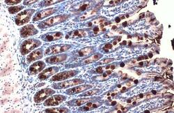
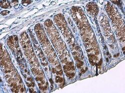
- MUC2 Polyclonal Antibody detects secreted MUC2 protein by immunohistochemical analysis. Sample: Paraffin-embedded mouse colon. MUC2 stained by MUC2 Polyclonal Antibody (Product # PA5-21329) diluted at 1:500. Antigen Retrieval: Citrate buffer, pH 6.0, 15 min.

- Submitted by
- Invitrogen Antibodies (provider)
- Main image
- Experimental details
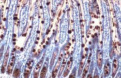
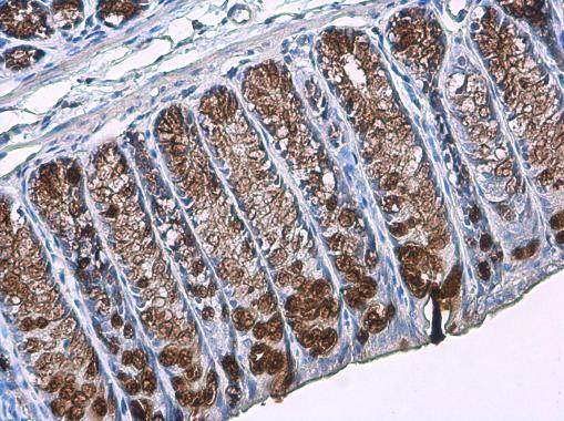
- MUC2 Polyclonal Antibody detects secreted MUC2 protein by immunohistochemical analysis. Sample: Paraffin-embedded mouse intestine. MUC2 stained by MUC2 Polyclonal Antibody (Product # PA5-21329) diluted at 1:500. Antigen Retrieval: Citrate buffer, pH 6.0, 15 min.

- Submitted by
- Invitrogen Antibodies (provider)
- Main image

- Experimental details
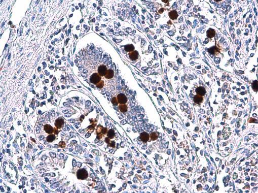
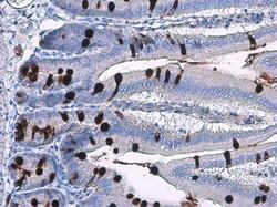
- Immunohistochemistry (Paraffin) analysis of MUC2 was performed in paraffin-embedded human intestine tissue using MUC2 Polyclonal Antibody (Product # PA5-21329) at a dilution of 1:750.
- Submitted by
- Invitrogen Antibodies (provider)
- Main image
- Experimental details
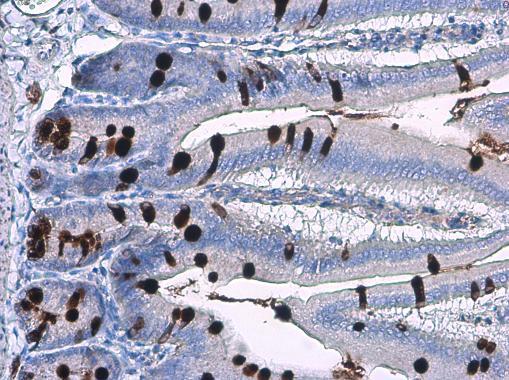
- Immunohistochemistry (Paraffin) analysis of MUC2 was performed in paraffin-embedded mouse colon tissue using MUC2 Polyclonal Antibody (Product # PA5-21329) at a dilution of 1:2000.
- Submitted by
- Invitrogen Antibodies (provider)
- Main image
- Experimental details
- Immunohistochemistry (Paraffin) analysis of MUC2 was performed in paraffin-embedded mouse intestine tissue using MUC2 Polyclonal Antibody (Product # PA5-21329) at a dilution of 1:2000.
Supportive validation
- Submitted by
- Invitrogen Antibodies (provider)
- Main image

- Experimental details
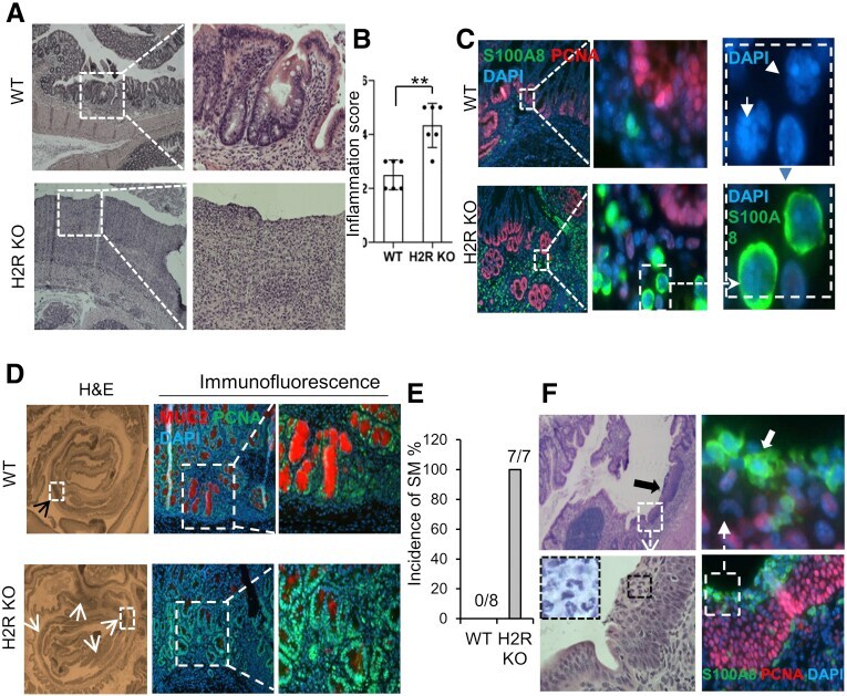
- Figure 2 H2R inactivation in mice resulted in aggravated DSS-induced colitis. ( A ) H&E staining (magnification: 40x and 200x) of middle and distal colon of WT and H2R-deficient mice after DSS treatment. Crypt polarity and architecture were disrupted in the middle and distal colon of H2R-deficient mice. ( B ) Inflammation scores were calculated based on the epithelial damage and infiltrating inflammatory cells from proximal to distal colon in 3 H&E sections from each mouse (total, 6 pairs of mice). Values are means +- SD. ** P < .01. ( C ) Immunofluorescence (magnification: 40x, 200x, and 1000x) of S100A8 (green), proliferating cell nuclear antigen (PCNA) (red), and 4',6-diamidino-2-phenylindole (DAPI) (blue, nuclear DNA staining) in the DSS-treated colon in WT and H2R KO mice. ( D ) DSS-induced hyperplasia ( top , indicated by a black arrowhead ) and dysplasia ( bottom , indicated by white arrowheads ) (magnification: 40x) in the colons of WT and H2R KO mice, respectively. Immunofluorescence (magnification: 200x and 400x) of MUC2 (red) and PCNA (green) shows much fewer MUC2-positive mature goblet cells in the DSS-treated colon of H2R KO mice. ( E ) The incidence of squamous metaplasia of DSS-treated WT (n = 8) and H2R KO (n = 7) mice. ( F ) H&E staining (magnification: 40x and 200x) and immunofluorescence (magnification: 200x and 1000x) of S100A8 (green, indicated by a white arrow) and PCNA (red) for the DSS-induced SM lesions (indicated by a black arrow ) in the distal colo