Explore
 Validate
 Learn
LearnPA1-32129
antibody from Invitrogen Antibodies
Targeting: ABCA1
ABC1, HDLDT1, TGD
 Western blot
 ELISA Immunocytochemistry Immunoprecipitation Immunohistochemistry Flow cytometry Gel shift Chromatin Immunoprecipitation
Antibody data
- Antibody Data
- Antigen structure
- References [0]
- Comments [0]
- Validations
- Western blot [4]
- Immunocytochemistry [4]
- Immunohistochemistry [1]
- Flow cytometry [2]
Submit
Validation data
Reference
Comment
Report error
- Product number
- PA1-32129 - Provider product page

- Provider
- Invitrogen Antibodies
- Product name
- ABCA1 Polyclonal Antibody
- Antibody type
- Polyclonal
- Antigen
- Synthetic peptide
- Description
- Store product as a concentrated solution. Centrifuge briefly prior to opening the vial.
- Reactivity
- Human, Mouse, Rat, Canine, Chicken/Avian, Hamster, Porcine
- Host
- Rabbit
- Isotype
- IgG
- Vial size
- 100 μL
- Concentration
- 1 mg/mL
- Storage
- Store at 4°C short term. For long term storage, store at -20°C, avoiding freeze/thaw cycles.
No comments: Submit comment
Supportive validation
- Submitted by
- Invitrogen Antibodies (provider)
- Main image

- Experimental details
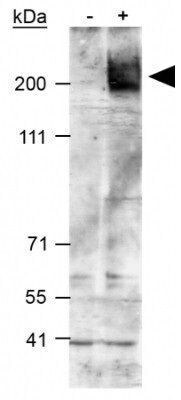
- Western Blot analysis of RAW264.7 cells treated with vehicle (-) or 9-cisretinoic acid and 22rhydroxycholesterol (+) using ABCA1 Polyclonal Antibody (Product # PA1-32129). Loading: 40 µg.
- Submitted by
- Invitrogen Antibodies (provider)
- Main image
- Experimental details
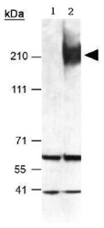
- Western Blot analysis of mouse peritoneal macrophages using ABCA1 Polyclonal Antibody (Product # PA1-32129). 1: T09 cell lysate. 2: Induced T09 cell lysate.

- Submitted by
- Invitrogen Antibodies (provider)
- Main image
- Experimental details
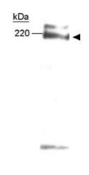
- Western Blot analysis of post-nuclear lysate of raw macrophages treated with 9-cisretinoic acid and 22R-hydroxycholesterol using ABCA1 Polyclonal Antibody (Product # PA1-32129). Loading: 40 µg.

- Submitted by
- Invitrogen Antibodies (provider)
- Main image
- Experimental details
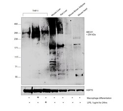
- Western blot was performed using anti-ABCA1 Polyclonal Antibody (Product # PA1-32129) and a 254 kDa band corresponding to ABCA1 was increased upon THP-1 differentiated to macrophages and LPS treatment in THP-1. Also, the expression was observed to be high in Mouse and Rat Liver when compared to other tissues. Few uncharacterized bands were also observed across the samples tested. Membrane enriched extracts (30 µg lysate) of THP-1 (Lane 1), THP-1 differentiated to macrophages (Lane 2), THP-1 treated with LPS (1 µg/mL for 24hrs) (Lane 3), Mouse Liver (Lane 4), Rat Liver (Lane 5), Mouse Brown Adipose (Lane 6) and Mouse Heart (Lane 7) were electrophoresed using NuPAGE™ 3-8% Tris-Acetate Protein Gels (Product # EA0378BOX). Resolved proteins were then transferred onto a nitrocellulose membrane (Product # IB23001) by iBlot® 2 Dry Blotting System (Product # IB21001). The blot was probed with the primary antibody (1:1000 dilution) and detected by chemiluminescence with Goat anti-Rabbit IgG (Heavy Chain), Superclonal™ Recombinant Secondary Antibody, HRP (Product # A27036, 1:4000 dilution) using the iBright FL 1000 (Product # A32752). Chemiluminescent detection was performed using SuperSignal™ West Dura Extended Duration Substrate (Product # 34076).
Supportive validation
- Submitted by
- Invitrogen Antibodies (provider)
- Main image

- Experimental details
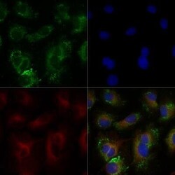
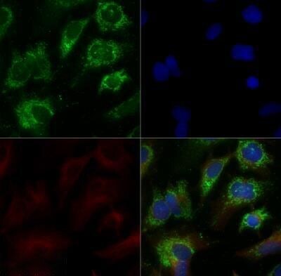
- Immunocytochemistry-Immunofluorescence analysis of ABCA1 in HepG2 cells. Cells were fixed with 10% formalin, permeabilized with 0.5% Triton-X100 and ABCA1 Polyclonal Antibody (Product # PA1-32129) (Green) at a dilution of 5 µg/mL was used as a primary antibody. Red : Tubulin. Blue: DAPI.

- Submitted by
- Invitrogen Antibodies (provider)
- Main image

- Experimental details
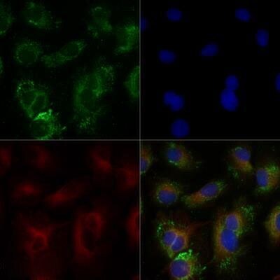
- Immunocytochemistry-Immunofluorescence analysis of ABCA1 in HepG2 cells(serum starvation 24hrs and treated with 1uM TO9). Cells were fixed with 10% formalin, permeabilized with 0.5% Triton-X100 and ABCA1 Polyclonal Antibody (Product # PA1-32129) (Green) at a dilution of 5 µg/mL was used as a primary antibody. Red : Tubulin. Blue: DAPI.

- Submitted by
- Invitrogen Antibodies (provider)
- Main image
- Experimental details
- Immunocytochemistry-Immunofluorescence analysis of ABCA1 in HepG2 cells. Cells were fixed with 10% formalin, permeabilized with 0.5% Triton-X100 and ABCA1 Polyclonal Antibody (Product # PA1-32129) (Green) at a dilution of 5 µg/mL was used as a primary antibody. Red : Tubulin. Blue: DAPI.
- Submitted by
- Invitrogen Antibodies (provider)
- Main image

- Experimental details
- Immunocytochemistry-Immunofluorescence analysis of ABCA1 in HepG2 cells(serum starvation 24hrs and treated with 1uM TO9). Cells were fixed with 10% formalin, permeabilized with 0.5% Triton-X100 and ABCA1 Polyclonal Antibody (Product # PA1-32129) (Green) at a dilution of 5 µg/mL was used as a primary antibody. Red : Tubulin. Blue: DAPI.
Supportive validation
- Submitted by
- Invitrogen Antibodies (provider)
- Main image
- Experimental details
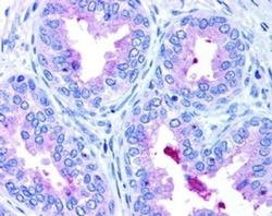
- Immunohistochemistry (Paraffin) analysis of ABCA1 in prostate tissue using ABCA1 Polyclonal Antibody (Product # PA1-32129).

Supportive validation
- Submitted by
- Invitrogen Antibodies (provider)
- Main image

- Experimental details
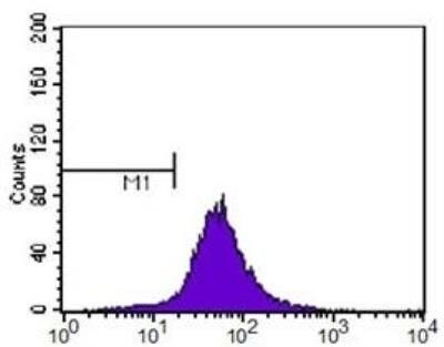
- Flow Cytometry analysis of Glypican 3 was performed in HeLa cells using Glypican 3 Monoclonal Antibody (GT764) (Product # MA5-31547) (Purple) at a dilution of 1:400. M1 is defined by unstained cells.

- Submitted by
- Invitrogen Antibodies (provider)
- Main image

- Experimental details
- Flow Cytometry analysis of Glypican 3 was performed in HeLa cells using Glypican 3 Monoclonal Antibody (GT764) (Product # MA5-31547) (Purple) at a dilution of 1:400. M1 is defined by unstained cells.