Explore
 Validate
 Learn
LearnPA1-16789
antibody from Invitrogen Antibodies
Targeting: ABCA1
ABC1, HDLDT1, TGD
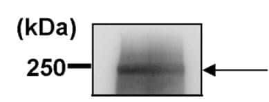
 Western blot
 Immunocytochemistry Immunoprecipitation Immunohistochemistry Flow cytometry
 Gel shift Chromatin Immunoprecipitation Other assay
Antibody data
- Antibody Data
- Antigen structure
- References [8]
- Comments [0]
- Validations
- Immunocytochemistry [5]
- Immunoprecipitation [1]
- Immunohistochemistry [1]
- Flow cytometry [2]
- Other assay [7]
Submit
Validation data
Reference
Comment
Report error
- Product number
- PA1-16789 - Provider product page

- Provider
- Invitrogen Antibodies
- Product name
- ABCA1 Polyclonal Antibody
- Antibody type
- Polyclonal
- Antigen
- Synthetic peptide
- Description
- ABCA1 is known to aggregate. DO NOT BOIL SAMPLES.
- Reactivity
- Human, Mouse, Rat, Canine, Chicken/Avian, Hamster, Porcine, Rabbit
- Host
- Rabbit
- Isotype
- IgG
- Vial size
- 100 μL
- Concentration
- 1 mg/mL
- Storage
- Store at 4°C short term. For long term storage, store at -20°C, avoiding freeze/thaw cycles.
Submitted references Givinostat-Liposomes: Anti-Tumor Effect on 2D and 3D Glioblastoma Models and Pharmacokinetics.
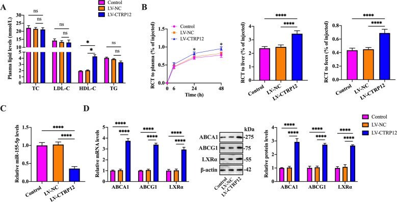
CTRP12 ameliorates atherosclerosis by promoting cholesterol efflux and inhibiting inflammatory response via the miR-155-5p/LXRα pathway.
The Liver X Receptor Is Upregulated in Monocyte-Derived Macrophages and Modulates Inflammatory Cytokines Based on LXRα Polymorphism.
ATP Binding Cassette Transporter A1 is Involved in Extracellular Secretion of Acetylated APE1/Ref-1.
PMP22 Regulates Cholesterol Trafficking and ABCA1-Mediated Cholesterol Efflux.
Calpains Released by T Lymphocytes Cleave TLR2 To Control IL-17 Expression.
Xanthine oxidoreductase is involved in macrophage foam cell formation and atherosclerosis development.
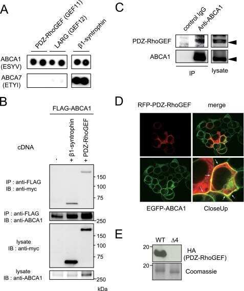
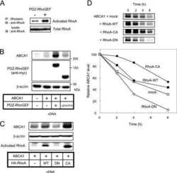
Binding of PDZ-RhoGEF to ATP-binding cassette transporter A1 (ABCA1) induces cholesterol efflux through RhoA activation and prevention of transporter degradation.
Taiarol L, Bigogno C, Sesana S, Kravicz M, Viale F, Pozzi E, Monza L, Carozzi VA, Meregalli C, Valtorta S, Moresco RM, Koch M, Barbugian F, Russo L, Dondio G, Steinkühler C, Re F
Cancers 2022 Jun 16;14(12)
Cancers 2022 Jun 16;14(12)
CTRP12 ameliorates atherosclerosis by promoting cholesterol efflux and inhibiting inflammatory response via the miR-155-5p/LXRα pathway.
Wang G, Chen JJ, Deng WY, Ren K, Yin SH, Yu XH
Cell death & disease 2021 Mar 10;12(3):254
Cell death & disease 2021 Mar 10;12(3):254
The Liver X Receptor Is Upregulated in Monocyte-Derived Macrophages and Modulates Inflammatory Cytokines Based on LXRα Polymorphism.
Kim HA, Baek WY, Han MH, Jung JY, Suh CH
Mediators of inflammation 2019;2019:6217548
Mediators of inflammation 2019;2019:6217548
ATP Binding Cassette Transporter A1 is Involved in Extracellular Secretion of Acetylated APE1/Ref-1.
Lee YR, Joo HK, Lee EO, Cho HS, Choi S, Kim CS, Jeon BH
International journal of molecular sciences 2019 Jun 28;20(13)
International journal of molecular sciences 2019 Jun 28;20(13)
PMP22 Regulates Cholesterol Trafficking and ABCA1-Mediated Cholesterol Efflux.
Zhou Y, Miles JR, Tavori H, Lin M, Khoshbouei H, Borchelt DR, Bazick H, Landreth GE, Lee S, Fazio S, Notterpek L
The Journal of neuroscience : the official journal of the Society for Neuroscience 2019 Jul 3;39(27):5404-5418
The Journal of neuroscience : the official journal of the Society for Neuroscience 2019 Jul 3;39(27):5404-5418
Calpains Released by T Lymphocytes Cleave TLR2 To Control IL-17 Expression.
Perez J, Dansou B, Hervé R, Levi C, Tamouza H, Vandermeersch S, Demey-Thomas E, Haymann JP, Zafrani L, Klatzmann D, Boissier MC, Letavernier E, Baud L
Journal of immunology (Baltimore, Md. : 1950) 2016 Jan 1;196(1):168-81
Journal of immunology (Baltimore, Md. : 1950) 2016 Jan 1;196(1):168-81
Xanthine oxidoreductase is involved in macrophage foam cell formation and atherosclerosis development.
Kushiyama A, Okubo H, Sakoda H, Kikuchi T, Fujishiro M, Sato H, Kushiyama S, Iwashita M, Nishimura F, Fukushima T, Nakatsu Y, Kamata H, Kawazu S, Higashi Y, Kurihara H, Asano T
Arteriosclerosis, thrombosis, and vascular biology 2012 Feb;32(2):291-8
Arteriosclerosis, thrombosis, and vascular biology 2012 Feb;32(2):291-8
Binding of PDZ-RhoGEF to ATP-binding cassette transporter A1 (ABCA1) induces cholesterol efflux through RhoA activation and prevention of transporter degradation.
Okuhira K, Fitzgerald ML, Tamehiro N, Ohoka N, Suzuki K, Sawada J, Naito M, Nishimaki-Mogami T
The Journal of biological chemistry 2010 May 21;285(21):16369-77
The Journal of biological chemistry 2010 May 21;285(21):16369-77
No comments: Submit comment
Supportive validation
- Submitted by
- Invitrogen Antibodies (provider)
- Main image
- Experimental details
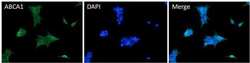
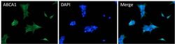
- Immunofluorescent analysis of ABCA1 (green) HEK293T cells. Cells fixed with 4% formaldehyde fixed cells were permeabilized and blocked with 1X PBS containing 5% BSA and 0.3% Triton X-100 for 1 hour at room temperature. Cells were probed with an ABCA1 polyclonal antibody (Product # PA1-16789) at a dilution of 1:100 overnight at 4°C in 1X PBS containing 1% BSA and 0.3% Triton X-100, washed with 1X PBS, and incubated with fluorophore-conjugated goat anti-rabbit IgG secondary antibody at a dilution of 1:200 for 1 hour at room temperature. Nuclei (blue) were stained with DAPI. Images were taken on a Leica DM1000 microscope at 40X magnification. Data courtesy of the Innovators Program.
- Submitted by
- Invitrogen Antibodies (provider)
- Main image
- Experimental details
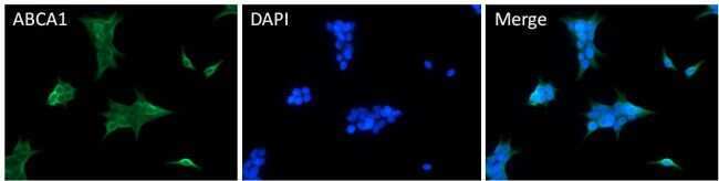
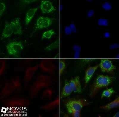
- Immunocytochemistry analysis of ABCA1 in HepG2 cells were grown to 60% confluency, serum starved for 24 hours, and then treated with 1 µM TO9 for 24 hours prior to being fixed for 10 minutes using 10% formalin and then permeabilized for 5 minutes using 1X TBS + 0.5% Triton-X100. Samples were incubated in ABCA1 polyclonal antibody (Product # PA1-16789) using a dilution of 5 µg/mL overnight at 4 °C followed by anti-rabbit DyLight 488 (Green) with a dilution of 1:500. Alpha tubulin (DM1A) was used as a co-stain at a 1:1000 dilution and detected with an anti-mouse DyLight 550 (Red) at a 1:500 dilution. Nuclei were counterstained with DAPI (Blue). Cells were imaged using a 40X objective.
- Submitted by
- Invitrogen Antibodies (provider)
- Main image

- Experimental details
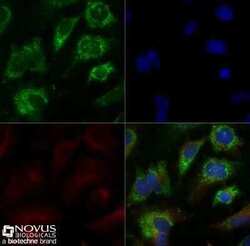
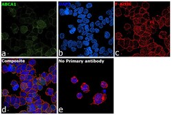
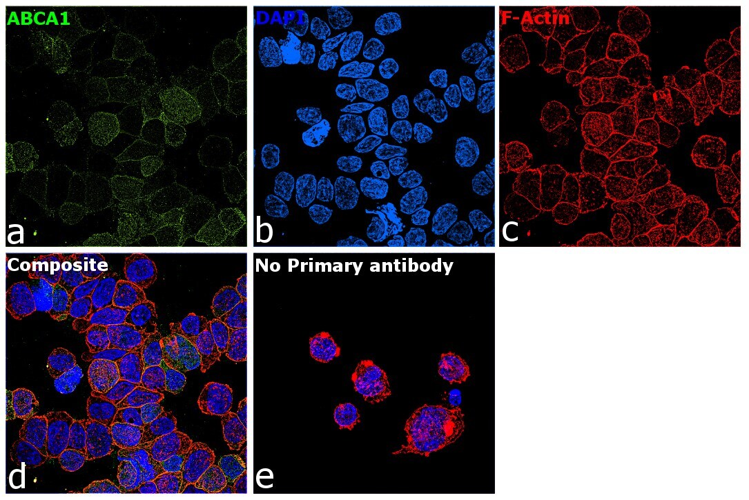
- Immunofluorescence analysis of ABCA1 was performed using log phase THP-1 cells. The cells were fixed with 4% paraformaldehyde for 10 minutes, permeabilized with 0.1% Triton™ X-100 for 15 minutes, and blocked with 2% BSA for 1 hour at room temperature. The cells were labeled with ABCA1 Polyclonal Antibody (3A1.891.3) (Product # PA1-16789) at 1:100 dilution in 0.1% BSA, incubated at 4 degree Celsius overnight and then with Donkey anti-Rabbit IgG (H+L) Highly Cross-Adsorbed Secondary Antibody, Alexa Fluor Plus 488 (Product # A32790) at a dilution of 1:2000 for 45 minutes at room temperature (Panel a: green). Nuclei (Panel b: blue) were stained with ProLong™ Diamond Antifade Mountant with DAPI (Product # P36962). F-actin (Panel c: red) was stained with Rhodamine Phalloidin (Product # R415, 1:300). Panel d represents the merged image showing membrane localization. Panel e represents control cells with no primary antibody to assess background. The images were captured at 60X magnification.

- Submitted by
- Invitrogen Antibodies (provider)
- Main image
- Experimental details
- Immunofluorescence analysis of ABCA1 was performed using log phase THP-1 cells. The cells were fixed with 4% paraformaldehyde for 10 minutes, permeabilized with 0.1% Triton™ X-100 for 15 minutes, and blocked with 2% BSA for 1 hour at room temperature. The cells were labeled with ABCA1 Polyclonal Antibody (3A1.891.3) (Product # PA1-16789) at 1:100 dilution in 0.1% BSA, incubated at 4 degree Celsius overnight and then with Donkey anti-Rabbit IgG (H+L) Highly Cross-Adsorbed Secondary Antibody, Alexa Fluor Plus 488 (Product # A32790) at a dilution of 1:2000 for 45 minutes at room temperature (Panel a: green). Nuclei (Panel b: blue) were stained with ProLong™ Diamond Antifade Mountant with DAPI (Product # P36962). F-actin (Panel c: red) was stained with Rhodamine Phalloidin (Product # R415, 1:300). Panel d represents the merged image showing membrane localization. Panel e represents control cells with no primary antibody to assess background. The images were captured at 60X magnification.
- Submitted by
- Invitrogen Antibodies (provider)
- Main image
- Experimental details
- Immunofluorescent analysis of ABCA1 (green) HEK293T cells. Cells fixed with 4% formaldehyde fixed cells were permeabilized and blocked with 1X PBS containing 5% BSA and 0.3% Triton X-100 for 1 hour at room temperature. Cells were probed with an ABCA1 polyclonal antibody (Product # PA1-16789) at a dilution of 1:100 overnight at 4°C in 1X PBS containing 1% BSA and 0.3% Triton X-100, washed with 1X PBS, and incubated with fluorophore-conjugated goat anti-rabbit IgG secondary antibody at a dilution of 1:200 for 1 hour at room temperature. Nuclei (blue) were stained with DAPI. Images were taken on a Leica DM1000 microscope at 40X magnification. Data courtesy of the Innovators Program.

Supportive validation
- Submitted by
- Invitrogen Antibodies (provider)
- Main image

- Experimental details
- Immunoprecipitation of ABCA1 was performed on THP-1 cells. Antigen-antibody complexes were formed by incubating 400 µg of THP-1 whole cell lysate (in 400 µL volume) with 4 µL of an ABCA1 polyclonal antibody (Product # PA1-16789) overnight at 4°C. The immune complexes were captured on 30 µL of protein G agarose, washed extensively, and eluted with 6X non-reducing Laemmli buffer. Samples were resolved on an 8% SDS-PAGE gel, transferred to a PVDF membrane, and blocked with 5% milk in TBST for 1 hour at room temperature. The membrane was probed with an ABCA1 polyclonal antibody (Product # PA1-16789) at a dilution of 1:500 for overnight at 4°C, washed in TBST, and probed with an HRP-conjugated goat anti-rabbit IgG secondary antibody at a dilution of 1:40,000 for 1 hour at room temperature. Chemiluminescent detection was performed using ECL substrate. Data courtesy of the Innovators Program.

Supportive validation
- Submitted by
- Invitrogen Antibodies (provider)
- Main image
- Experimental details
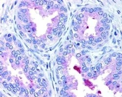
- Immunohistochemical analysis of ABCA1 in human prostate epithelium showing luminal and membrane staining. Samples were incubated in ABCA1 polyclonal antibody (Product # PA1-16789).

Supportive validation
- Submitted by
- Invitrogen Antibodies (provider)
- Main image

- Experimental details
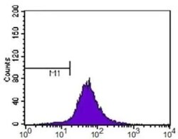
- Flow cytometry of ABCA1 in HeLa cells. Samples were incubated in ABCA1 polyclonal antibody (Product # PA1-16789) using a dilution of 1:400. Alexa Fluor 488 secondary (shown in purple). M1 is defined by unstained cells.

- Submitted by
- Invitrogen Antibodies (provider)
- Main image
- Experimental details
- Flow cytometry of ABCA1 in HeLa cells. Samples were incubated in ABCA1 polyclonal antibody (Product # PA1-16789) using a dilution of 1:400. Alexa Fluor 488 secondary (shown in purple). M1 is defined by unstained cells.

Supportive validation
- Submitted by
- Invitrogen Antibodies (provider)
- Main image

- Experimental details
- NULL
- Submitted by
- Invitrogen Antibodies (provider)
- Main image
- Experimental details
- NULL

- Submitted by
- Invitrogen Antibodies (provider)
- Main image

- Experimental details
- Immunoprecipitation of ABCA1 was performed on THP-1 cells. Antigen-antibody complexes were formed by incubating 400 µg of THP-1 whole cell lysate (in 400 µL volume) with 4 µL of an ABCA1 polyclonal antibody (Product # PA1-16789) overnight at 4°C. The immune complexes were captured on 30 µL of protein G agarose, washed extensively, and eluted with 6X non-reducing Laemmli buffer. Samples were resolved on an 8% SDS-PAGE gel, transferred to a PVDF membrane, and blocked with 5% milk in TBST for 1 hour at room temperature. The membrane was probed with an ABCA1 polyclonal antibody (Product # PA1-16789) at a dilution of 1:500 for overnight at 4øC, washed in TBST, and probed with an HRP-conjugated goat anti-rabbit IgG secondary antibody at a dilution of 1:40,000 for 1 hour at room temperature. Chemiluminescent detection was performed using ECL substrate. Data courtesy of the Innovators Program.
- Submitted by
- Invitrogen Antibodies (provider)
- Main image

- Experimental details
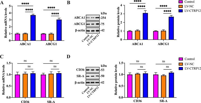
- Fig. 2 Effects of CTRP12 on the expression of cholesterol efflux and uptake markers. A - D THP-1 macrophages were incubated with 50 ug/mL ox-LDL for 48 h, followed by treatment with PBS, LV-NC, or LV-CTRP12 for 72 h. A , B The expression of ABCA1 and ABCG1 was determined by qRT-PCR and western blot. C , D Detection of CD36 and SR-A expression using qRT-PCR and western blot. Data are expressed as the mean +- SD from three independent experiments. **** P < 0.0001; ns not significant.
- Submitted by
- Invitrogen Antibodies (provider)
- Main image
- Experimental details
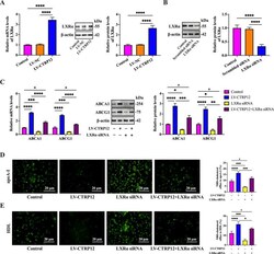
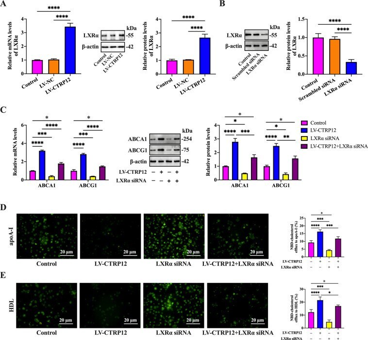
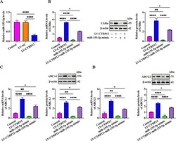
- Fig. 3 Involvement of LXRalpha in CTRP12-induced upregulation of ABCA1 and ABCG1 expression. A THP-1 macrophages were treated with 50 ug/mL ox-LDL for 48 h, followed by transfection with PBS, LV-NC, or LV-CTRP12 for 72 h. LXRalpha expression was determined by qRT-PCR and western blot. B After transfection with scrambled siRNA or LXRalpha siRNA for 48 h, cell lysates were immunoblotted with indicated antibodies. C - E THP-1 macrophages were loaded with 50 ug/mL ox-LDL for 48 h, transfected with LXRalpha siRNA for another 48 h and then treated with LV-CTRP12 for 72 h. C The mRNA and protein levels of ABCA1 and ABCG1 were detected by qRT-PCR and western blot, respectively. D , E Representative fluorescent images of NBD-cholesterol burden (x200) and quantitative analyses of cholesterol efflux to apoA-I and HDL. Scale bar = 20 mum. Data are expressed as the mean +- SD from three independent experiments. * P < 0.05, ** P < 0.01, *** P < 0.001, **** P < 0.0001.
- Submitted by
- Invitrogen Antibodies (provider)
- Main image
- Experimental details
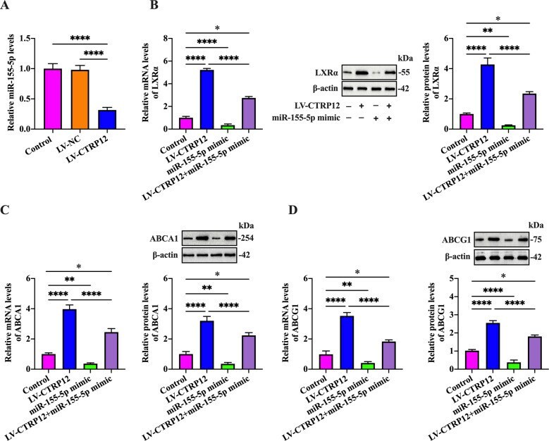
- Fig. 5 CTRP12-induced upregulation of LXRalpha, ABCA1, and ABCG1 is mediated by miR-155-5p. A THP-1 macrophages were treated with 50 ug/mL ox-LDL for 48 h and then transfected with PBS, LV-NC, or LV-CTRP12 for 72 h, followed by detection of miR-155-5p expression using qRT-PCR. B - D THP-1 macrophages were loaded with 50 ug/mL ox-LDL for 48 h, transfected with miR-155-5p mimic for another 48 h and then transduced with LV-CTRP12 for 72 h. The mRNA and protein levels of LXRalpha, ABCA1, and ABCG1 were detected by qRT-PCR and western blot, respectively. Data are represented as the mean +- SD from three independent experiments. * P < 0.05, ** P < 0.01, **** P < 0.0001.
- Submitted by
- Invitrogen Antibodies (provider)
- Main image
- Experimental details
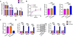
- Fig. 8 Effects of CTRP12 on plasma HDL-C levels and RCT in apoE -/- mice. A Plasma levels of TC, TG, HDL-C, and LDL-C were determined using the commercial kits ( n = 10). B Mice were injected intraperitoneally with [ 3 H]-cholesterol-labeled J774 macrophages. The radioactivity in the plasma, liver, and feces were assessed by a liquid scintillation counter ( n = 5). C , D Detection of miR-155-5p, LXRalpha, ABCA1, and ABCG1 expression in the aortas by qRT-PCR and western blot ( n = 10). Data are represented as the mean +- SD. * P < 0.05, **** P < 0.0001; ns not significant.