Explore
 Validate
 Learn
Learn Western blot
 ELISA
ELISAAntibody data
- Antibody Data
- Antigen structure
- References [46]
- Comments [0]
- Validations
- Western blot [1]
- Immunocytochemistry [1]
- Immunohistochemistry [2]
Submit
Validation data
Reference
Comment
Report error
- Product number
- 16675-1-AP - Provider product page

- Provider
- Proteintech Group
- Proper citation
- Proteintech Cat#16675-1-AP, RRID:AB_2226463
- Product name
- AMFR antibody
- Antibody type
- Polyclonal
- Description
- KD/KO validated AMFR antibody (Cat. #16675-1-AP) is a rabbit polyclonal antibody that shows reactivity with human, mouse, rat and has been validated for the following applications: IF, IHC, IP, WB,ELISA.
- Reactivity
- Human, Mouse, Rat
- Host
- Rabbit
- Conjugate
- Unconjugated
- Isotype
- IgG
- Vial size
- 20ul, 150ul
Submitted references Nur77 attenuates Paneth cell necroptosis-induced intestinal inflammation through regulating endoplasmic reticulum homeostasis in sepsis.
O-GlcNAcylation of AMFR stabilizes TSPAN4 to regulate migrasome formation for viral release.
eIF3f promotes tumour malignancy by remodelling fatty acid biosynthesis in hepatocellular carcinoma.
ER-phagy Activation by AMFR Attenuates Cardiac Fibrosis Post-Myocardial Infarction via mTORC1 Pathway.
AMFR-mediated ER-phagy regulation and therapeutic targeting in osteosarcoma: a multifunctional nanoplatform strategy.
Dysregulation of the SREBP pathway is associated with poor prognosis and serves as a potential biomarker for the diagnosis of hepatocellular carcinoma.
Knockdown of AMFR Alleviates Atrial Fibrosis in Atrial Fibrillation by Stabilizing SOD1 Protein Expression.
UFMylation of BiP/GRP78 Is Crucial for Maintenance of Endoplasmic Reticulum Homeostasis.
Cannabidiol Enhances the Anticancer Activity of Etoposide on Prostate Cancer Cells.
The autocrine motility factor receptor delays the pathological progression of Alzheimer's disease via regulating the ubiquitination-mediated degradation of APP.
The p97-UBXD8 complex maintains peroxisome abundance by suppressing pexophagy.
gp78-regulated KAP1 phosphorylation induces radioresistance in breast cancer by facilitating PPP1CC/PPP2CA ubiquitination.
AMFR-mediated Flavivirus NS2A ubiquitination subverts ER-phagy to augment viral pathogenicity.
Autocrine motility factor receptor promotes the malignancy of glioblastoma by regulating cell migration and invasion.
New mode of action of curcumin on prostate cancer cells: Modulation of endoplasmic reticulum-associated degradation mechanism and estrogenic signaling.
Standigm ASK™: knowledge graph and artificial intelligence platform applied to target discovery in idiopathic pulmonary fibrosis.
Nicotine-derived NNK promotes CRC progression through activating TMUB1/AKT pathway in METTL14/YTHDF2-mediated m6A manner.
Circadian Oscillation Pattern of Endoplasmic Reticulum Quality Control (ERQC) Components in Human Embryonic Kidney HEK293 Cells.
Estrogens drive the endoplasmic reticulum-associated degradation and promote proto-oncogene c-Myc expression in prostate cancer cells by androgen receptor/estrogen receptor signaling.
Triiodothyronine positively regulates endoplasmic reticulum-associated degradation (ERAD) and promotes androgenic signaling in androgen-dependent prostate cancer cells.
Suberoylanilide hydroxamic acid upregulates reticulophagy receptor expression and promotes cell death in hepatocellular carcinoma cells.
1,25(OH)(2) D(3) induced vitamin D receptor signaling negatively regulates endoplasmic reticulum-associated degradation (ERAD) and androgen receptor signaling in human prostate cancer cells.
Heteromeric clusters of ubiquitinated ER-shaping proteins drive ER-phagy.
Ubiquitination regulates ER-phagy and remodelling of endoplasmic reticulum.
Dexamethasone-stimulated glucocorticoid receptor signaling positively regulates the endoplasmic reticulum-associated degradation (ERAD) mechanism in hepatocellular carcinoma cells.
The chemokine CCL1 facilitates pulmonary fibrosis by promoting macrophage migration and M2 polarization.
Progesterone regulates the endoplasmic reticulum-associated degradation and Unfolded Protein Response axis by mimicking the androgenic stimulation in prostate cancer cells.
The p97-UBXD8 complex regulates ER-Mitochondria contact sites by altering membrane lipid saturation and composition.
Gp78 deficiency in hepatocytes alleviates hepatic ischemia-reperfusion injury via suppressing ACSL4-mediated ferroptosis.
3β-hydroxysteroid-Δ24 reductase dampens anti-viral innate immune responses by targeting K27 ubiquitination of MAVS and STING.
The GR-gp78 Pathway is involved in Hepatic Lipid Accumulation Induced by Overexpression of 11β-HSD1.
Regulation of membrane fluidity by RNF145-triggered degradation of the lipid hydrolase ADIPOR2.
Basal Gp78-dependent mitophagy promotes mitochondrial health and limits mitochondrial ROS.
AAA-ATPase valosin-containing protein binds the transcription factor SREBP1 and promotes its proteolytic activation by rhomboid protease RHBDL4.
Ubiquitination of NLRP3 by gp78/Insig-1 restrains NLRP3 inflammasome activation.
Pharmacological induction of AMFR increases functional EAAT2 oligomer levels and reduces epileptic seizures in mice.
ERAD components Derlin-1 and Derlin-2 are essential for postnatal brain development and motor function.
Spermine Protects Cardiomyocytes from High Glucose-Induced Energy Disturbance by Targeting the CaSR-gp78-Ubiquitin Proteasome System.
Reticulon and CLIMP-63 regulate nanodomain organization of peripheral ER tubules.
MARCH6 and TRC8 facilitate the quality control of cytosolic and tail-anchored proteins.
The sterol-responsive RNF145 E3 ubiquitin ligase mediates the degradation of HMG-CoA reductase together with gp78 and Hrd1.
iRhom2 is essential for innate immunity to RNA virus by antagonizing ER- and mitochondria-associated degradation of VISA.
Calcium sensing receptor protects high glucose-induced energy metabolism disorder via blocking gp78-ubiquitin proteasome pathway.
Aberrant corticosteroid metabolism in tumor cells enables GR takeover in enzalutamide resistant prostate cancer.
BOK Is a Non-canonical BCL-2 Family Effector of Apoptosis Regulated by ER-Associated Degradation.
The E3 ubiquitin ligase AMFR and INSIG1 bridge the activation of TBK1 kinase by modifying the adaptor STING.
Cui C, Huo Q, Ran S, Wang W, Wei H, Peng J
Journal of advanced research 2026 May;83:269-285
Journal of advanced research 2026 May;83:269-285
O-GlcNAcylation of AMFR stabilizes TSPAN4 to regulate migrasome formation for viral release.
Yu L, Li J, Han Y, Yang X, Fu Y, Zhang W, Jiu Y, Cheng L, Ding B
Nature communications 2026 Jan 7;17(1):1506
Nature communications 2026 Jan 7;17(1):1506
eIF3f promotes tumour malignancy by remodelling fatty acid biosynthesis in hepatocellular carcinoma.
Zhou S, Zhang L, You Y, Yu K, Tie X, Gao Y, Chen Y, Yao F, Zhang R, Hao X, Fang C, Li X, Li Q, Wang X
Journal of hepatology 2025 Sep;83(3):712-728
Journal of hepatology 2025 Sep;83(3):712-728
ER-phagy Activation by AMFR Attenuates Cardiac Fibrosis Post-Myocardial Infarction via mTORC1 Pathway.
Wang Z, Niu K, Liu W, Wang X, Yang B, Li T, Chen Y, Jin Y, Chen Y, Lin Y, Jin X
Advanced science (Weinheim, Baden-Wurttemberg, Germany) 2025 Oct;12(37):e04552
Advanced science (Weinheim, Baden-Wurttemberg, Germany) 2025 Oct;12(37):e04552
AMFR-mediated ER-phagy regulation and therapeutic targeting in osteosarcoma: a multifunctional nanoplatform strategy.
Zhao Q, Lu X, Xu T, Gao Z, Peng L, Zhu B, Wang W, Liu Z, Yang G, Zhao H, Song Z, Lou Q, Li J, Ren Z, Yu Z, Jesus M F, Cui D
Journal of nanobiotechnology 2025 Nov 18;23(1):717
Journal of nanobiotechnology 2025 Nov 18;23(1):717
Dysregulation of the SREBP pathway is associated with poor prognosis and serves as a potential biomarker for the diagnosis of hepatocellular carcinoma.
Li X, Wang Y, Liu J, Gao T, Cao L, Yan M, Li N
Molecular medicine reports 2025 May;31(5)
Molecular medicine reports 2025 May;31(5)
Knockdown of AMFR Alleviates Atrial Fibrosis in Atrial Fibrillation by Stabilizing SOD1 Protein Expression.
Geng S, Ruan Z, Ying L, Liu Z
Journal of cardiovascular pharmacology and therapeutics 2025 Jan-Dec;30:10742484251356140
Journal of cardiovascular pharmacology and therapeutics 2025 Jan-Dec;30:10742484251356140
UFMylation of BiP/GRP78 Is Crucial for Maintenance of Endoplasmic Reticulum Homeostasis.
Li Z, Liang Q, Guo Y, Wang Y, Wang M, Cong YS
FASEB journal : official publication of the Federation of American Societies for Experimental Biology 2025 Aug 15;39(15):e70905
FASEB journal : official publication of the Federation of American Societies for Experimental Biology 2025 Aug 15;39(15):e70905
Cannabidiol Enhances the Anticancer Activity of Etoposide on Prostate Cancer Cells.
Erzurumlu Y, Catakli D
Cannabis and cannabinoid research 2025 Apr;10(2):258-276
Cannabis and cannabinoid research 2025 Apr;10(2):258-276
The autocrine motility factor receptor delays the pathological progression of Alzheimer's disease via regulating the ubiquitination-mediated degradation of APP.
Zhang J, Liu C, Liu J, Cui Y, Hou Y, Song Q, Zhang X, Wang X, Zhang Q, Cao M, Wang W, Wang P, Wang Y
Alzheimer's research & therapy 2025 Apr 29;17(1):95
Alzheimer's research & therapy 2025 Apr 29;17(1):95
The p97-UBXD8 complex maintains peroxisome abundance by suppressing pexophagy.
Montes ID, Amirthagunanathan S, Joshi AS, Raman M
bioRxiv : the preprint server for biology 2024 Sep 26;
bioRxiv : the preprint server for biology 2024 Sep 26;
gp78-regulated KAP1 phosphorylation induces radioresistance in breast cancer by facilitating PPP1CC/PPP2CA ubiquitination.
Han Y, Xiao M, Zhao S, Wang H, Li R, Xu B
iScience 2024 Sep 20;27(9):110847
iScience 2024 Sep 20;27(9):110847
AMFR-mediated Flavivirus NS2A ubiquitination subverts ER-phagy to augment viral pathogenicity.
Zhang L, Wang H, Han C, Dong Q, Yan J, Guo W, Shan C, Zhao W, Chen P, Huang R, Wu Y, Chen Y, Qin Y, Chen M
Nature communications 2024 Nov 6;15(1):9578
Nature communications 2024 Nov 6;15(1):9578
Autocrine motility factor receptor promotes the malignancy of glioblastoma by regulating cell migration and invasion.
Zhang Y, Wang X, Chen G, Lu Y, Chen Q
Neurological research 2024 Jan;46(1):89-97
Neurological research 2024 Jan;46(1):89-97
New mode of action of curcumin on prostate cancer cells: Modulation of endoplasmic reticulum-associated degradation mechanism and estrogenic signaling.
Erzurumlu Y, Dogan HK, Catakli D
Journal of biochemical and molecular toxicology 2024 Jan;38(1):e23636
Journal of biochemical and molecular toxicology 2024 Jan;38(1):e23636
Standigm ASK™: knowledge graph and artificial intelligence platform applied to target discovery in idiopathic pulmonary fibrosis.
Han S, Lee JE, Kang S, So M, Jin H, Lee JH, Baek S, Jun H, Kim TY, Lee YS
Briefings in bioinformatics 2024 Jan 22;25(2)
Briefings in bioinformatics 2024 Jan 22;25(2)
Nicotine-derived NNK promotes CRC progression through activating TMUB1/AKT pathway in METTL14/YTHDF2-mediated m6A manner.
Jiang M, Han J, Ma Q, Chen X, Xu R, Wang Q, Zheng J, Wang W, Song J, Huang Y, Chen Y
Journal of hazardous materials 2024 Apr 5;467:133692
Journal of hazardous materials 2024 Apr 5;467:133692
Circadian Oscillation Pattern of Endoplasmic Reticulum Quality Control (ERQC) Components in Human Embryonic Kidney HEK293 Cells.
Erzurumlu Y, Catakli D, Dogan HK
Journal of circadian rhythms 2023;21:1
Journal of circadian rhythms 2023;21:1
Estrogens drive the endoplasmic reticulum-associated degradation and promote proto-oncogene c-Myc expression in prostate cancer cells by androgen receptor/estrogen receptor signaling.
Erzurumlu Y, Dogan HK, Catakli D, Aydogdu E, Muhammed MT
Journal of cell communication and signaling 2023 Sep;17(3):793-811
Journal of cell communication and signaling 2023 Sep;17(3):793-811
Triiodothyronine positively regulates endoplasmic reticulum-associated degradation (ERAD) and promotes androgenic signaling in androgen-dependent prostate cancer cells.
Erzurumlu Y, Muhammed MT
Cellular signalling 2023 Sep;109:110745
Cellular signalling 2023 Sep;109:110745
Suberoylanilide hydroxamic acid upregulates reticulophagy receptor expression and promotes cell death in hepatocellular carcinoma cells.
Li JY, Tian T, Han B, Yang T, Guo YX, Wu JY, Chen YS, Yang Q, Xie RJ
World journal of gastroenterology 2023 Sep 14;29(34):5038-5053
World journal of gastroenterology 2023 Sep 14;29(34):5038-5053
1,25(OH)(2) D(3) induced vitamin D receptor signaling negatively regulates endoplasmic reticulum-associated degradation (ERAD) and androgen receptor signaling in human prostate cancer cells.
Erzurumlu Y, Aydogdu E, Dogan HK, Catakli D, Muhammed MT, Buyuksandic B
Cellular signalling 2023 Mar;103:110577
Cellular signalling 2023 Mar;103:110577
Heteromeric clusters of ubiquitinated ER-shaping proteins drive ER-phagy.
Foronda H, Fu Y, Covarrubias-Pinto A, Bocker HT, González A, Seemann E, Franzka P, Bock A, Bhaskara RM, Liebmann L, Hoffmann ME, Katona I, Koch N, Weis J, Kurth I, Gleeson JG, Reggiori F, Hummer G, Kessels MM, Qualmann B, Mari M, Dikić I, Hübner CA
Nature 2023 Jun;618(7964):402-410
Nature 2023 Jun;618(7964):402-410
Ubiquitination regulates ER-phagy and remodelling of endoplasmic reticulum.
González A, Covarrubias-Pinto A, Bhaskara RM, Glogger M, Kuncha SK, Xavier A, Seemann E, Misra M, Hoffmann ME, Bräuning B, Balakrishnan A, Qualmann B, Dötsch V, Schulman BA, Kessels MM, Hübner CA, Heilemann M, Hummer G, Dikić I
Nature 2023 Jun;618(7964):394-401
Nature 2023 Jun;618(7964):394-401
Dexamethasone-stimulated glucocorticoid receptor signaling positively regulates the endoplasmic reticulum-associated degradation (ERAD) mechanism in hepatocellular carcinoma cells.
Erzurumlu Y, Dogan HK, Cataklı D
Steroids 2023 Jul;195:109238
Steroids 2023 Jul;195:109238
The chemokine CCL1 facilitates pulmonary fibrosis by promoting macrophage migration and M2 polarization.
Liu S, Zhang Z, Wang Y, Zhang Y, Min J, Li X, Liu S
International immunopharmacology 2023 Jul;120:110343
International immunopharmacology 2023 Jul;120:110343
Progesterone regulates the endoplasmic reticulum-associated degradation and Unfolded Protein Response axis by mimicking the androgenic stimulation in prostate cancer cells.
Erzurumlu Y, Dogan HK, Catakli D
Molecular biology reports 2023 Feb;50(2):1253-1265
Molecular biology reports 2023 Feb;50(2):1253-1265
The p97-UBXD8 complex regulates ER-Mitochondria contact sites by altering membrane lipid saturation and composition.
Ganji R, Paulo JA, Xi Y, Kline I, Zhu J, Clemen CS, Weihl CC, Purdy JG, Gygi SP, Raman M
Nature communications 2023 Feb 6;14(1):638
Nature communications 2023 Feb 6;14(1):638
Gp78 deficiency in hepatocytes alleviates hepatic ischemia-reperfusion injury via suppressing ACSL4-mediated ferroptosis.
Li C, Wu Y, Chen K, Chen R, Xu S, Yang B, Lian Z, Wang X, Wang K, Xie H, Zheng S, Liu Z, Wang D, Xu X
Cell death & disease 2023 Dec 8;14(12):810
Cell death & disease 2023 Dec 8;14(12):810
3β-hydroxysteroid-Δ24 reductase dampens anti-viral innate immune responses by targeting K27 ubiquitination of MAVS and STING.
Liu Q, Chen S, Tian R, Xue B, Li H, Guo M, Liu S, Yan M, You R, Wang L, Yang D, Wan M, Zhu H
Journal of virology 2023 Dec 21;97(12):e0151323
Journal of virology 2023 Dec 21;97(12):e0151323
The GR-gp78 Pathway is involved in Hepatic Lipid Accumulation Induced by Overexpression of 11β-HSD1.
Hu M, Han T, Pan Q, Ni D, Gao F, Wang L, Ren H, Zhang X, Jiao H, Wang Y, Dai D, Man Y, Tang W, Sun Y, Li W, Li J, Li G
International journal of biological sciences 2022;18(8):3107-3121
International journal of biological sciences 2022;18(8):3107-3121
Regulation of membrane fluidity by RNF145-triggered degradation of the lipid hydrolase ADIPOR2.
Volkmar N, Gawden-Bone CM, Williamson JC, Nixon-Abell J, West JA, St George-Hyslop PH, Kaser A, Lehner PJ
The EMBO journal 2022 Oct 4;41(19):e110777
The EMBO journal 2022 Oct 4;41(19):e110777
Basal Gp78-dependent mitophagy promotes mitochondrial health and limits mitochondrial ROS.
Alan P, Vandevoorde KR, Joshi B, Cardoen B, Gao G, Mohammadzadeh Y, Hamarneh G, Nabi IR
Cellular and molecular life sciences : CMLS 2022 Oct 25;79(11):565
Cellular and molecular life sciences : CMLS 2022 Oct 25;79(11):565
AAA-ATPase valosin-containing protein binds the transcription factor SREBP1 and promotes its proteolytic activation by rhomboid protease RHBDL4.
Shibuya K, Ebihara K, Ebihara C, Sawayama N, Isoda M, Yamamuro D, Takahashi M, Nagashima S, Ishibashi S
The Journal of biological chemistry 2022 Jun;298(6):101936
The Journal of biological chemistry 2022 Jun;298(6):101936
Ubiquitination of NLRP3 by gp78/Insig-1 restrains NLRP3 inflammasome activation.
Xu T, Yu W, Fang H, Wang Z, Chi Z, Guo X, Jiang D, Zhang K, Chen S, Li M, Guo Y, Zhang J, Yang D, Yu Q, Wang D, Zhang X
Cell death and differentiation 2022 Aug;29(8):1582-1595
Cell death and differentiation 2022 Aug;29(8):1582-1595
Pharmacological induction of AMFR increases functional EAAT2 oligomer levels and reduces epileptic seizures in mice.
Sha L, Li G, Zhang X, Lin Y, Qiu Y, Deng Y, Zhu W, Xu Q
JCI insight 2022 Aug 8;7(15)
JCI insight 2022 Aug 8;7(15)
ERAD components Derlin-1 and Derlin-2 are essential for postnatal brain development and motor function.
Sugiyama T, Murao N, Kadowaki H, Takao K, Miyakawa T, Matsushita Y, Katagiri T, Futatsugi A, Shinmyo Y, Kawasaki H, Sakai J, Shiomi K, Nakazato M, Takeda K, Mikoshiba K, Ploegh HL, Ichijo H, Nishitoh H
iScience 2021 Jul 23;24(7):102758
iScience 2021 Jul 23;24(7):102758
Spermine Protects Cardiomyocytes from High Glucose-Induced Energy Disturbance by Targeting the CaSR-gp78-Ubiquitin Proteasome System.
Wang Y, Wang Y, Li F, Zhang X, Li H, Yang G, Xu C, Wei C
Cardiovascular drugs and therapy 2021 Feb;35(1):73-85
Cardiovascular drugs and therapy 2021 Feb;35(1):73-85
Reticulon and CLIMP-63 regulate nanodomain organization of peripheral ER tubules.
Gao G, Zhu C, Liu E, Nabi IR
PLoS biology 2019 Aug;17(8):e3000355
PLoS biology 2019 Aug;17(8):e3000355
MARCH6 and TRC8 facilitate the quality control of cytosolic and tail-anchored proteins.
Stefanovic-Barrett S, Dickson AS, Burr SP, Williamson JC, Lobb IT, van den Boomen DJ, Lehner PJ, Nathan JA
EMBO reports 2018 May;19(5)
EMBO reports 2018 May;19(5)
The sterol-responsive RNF145 E3 ubiquitin ligase mediates the degradation of HMG-CoA reductase together with gp78 and Hrd1.
Menzies SA, Volkmar N, van den Boomen DJ, Timms RT, Dickson AS, Nathan JA, Lehner PJ
eLife 2018 Dec 13;7
eLife 2018 Dec 13;7
iRhom2 is essential for innate immunity to RNA virus by antagonizing ER- and mitochondria-associated degradation of VISA.
Luo WW, Li S, Li C, Zheng ZQ, Cao P, Tong Z, Lian H, Wang SY, Shu HB, Wang YY
PLoS pathogens 2017 Nov;13(11):e1006693
PLoS pathogens 2017 Nov;13(11):e1006693
Calcium sensing receptor protects high glucose-induced energy metabolism disorder via blocking gp78-ubiquitin proteasome pathway.
Wang Y, Gao P, Wei C, Li H, Zhang L, Zhao Y, Wu B, Tian Y, Zhang W, Wu L, Wang R, Xu C
Cell death & disease 2017 May 18;8(5):e2799
Cell death & disease 2017 May 18;8(5):e2799
Aberrant corticosteroid metabolism in tumor cells enables GR takeover in enzalutamide resistant prostate cancer.
Li J, Alyamani M, Zhang A, Chang KH, Berk M, Li Z, Zhu Z, Petro M, Magi-Galluzzi C, Taplin ME, Garcia JA, Courtney K, Klein EA, Sharifi N
eLife 2017 Feb 13;6
eLife 2017 Feb 13;6
BOK Is a Non-canonical BCL-2 Family Effector of Apoptosis Regulated by ER-Associated Degradation.
Llambi F, Wang YM, Victor B, Yang M, Schneider DM, Gingras S, Parsons MJ, Zheng JH, Brown SA, Pelletier S, Moldoveanu T, Chen T, Green DR
Cell 2016 Apr 7;165(2):421-33
Cell 2016 Apr 7;165(2):421-33
The E3 ubiquitin ligase AMFR and INSIG1 bridge the activation of TBK1 kinase by modifying the adaptor STING.
Wang Q, Liu X, Cui Y, Tang Y, Chen W, Li S, Yu H, Pan Y, Wang C
Immunity 2014 Dec 18;41(6):919-33
Immunity 2014 Dec 18;41(6):919-33
No comments: Submit comment
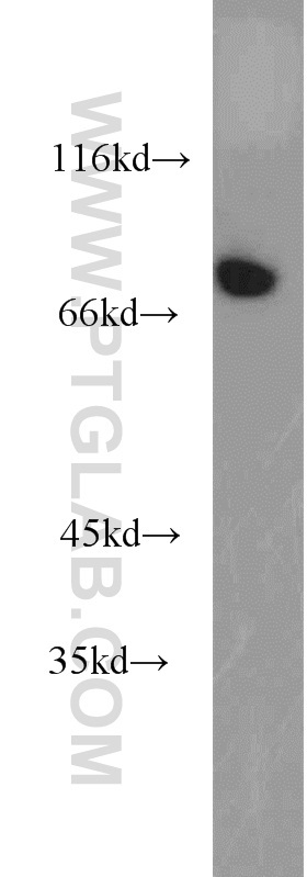
Supportive validation
- Submitted by
- Proteintech Group (provider)
- Main image

- Experimental details
- HepG2 cells were subjected to SDS PAGE followed by western blot with 16675-1-AP(AMFR antibody) at dilution of 1:800
- Sample type
- cell line
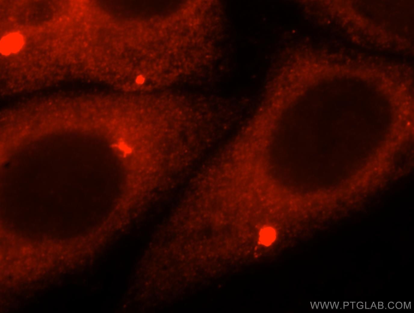
Supportive validation
- Submitted by
- Proteintech Group (provider)
- Main image
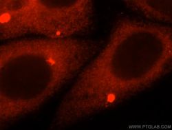
- Experimental details
- Immunofluorescent analysis of HepG2 cells, using AMFR antibody 16675-1-AP at 1:25 dilution and Rhodamine-labeled goat anti-rabbit IgG (red).
- Sample type
- cell line
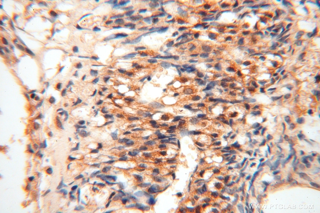
Supportive validation
- Submitted by
- Proteintech Group (provider)
- Main image
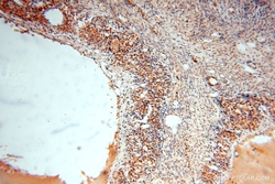
- Experimental details
- Immunohistochemical of paraffin-embedded human ovary using 16675-1-AP(AMFR antibody) at dilution of 1:100 (under 10x lens)
- Sample type
- tissue

- Submitted by
- Proteintech Group (provider)
- Main image
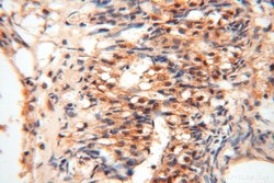
- Experimental details
- Immunohistochemical of paraffin-embedded human ovary using 16675-1-AP(AMFR antibody) at dilution of 1:100 (under 40x lens)
- Sample type
- tissue