Explore
 Validate
 Learn
Learn Western blot
 Immunocytochemistry
Antibody data
- Antibody Data
- Antigen structure
- References [0]
- Comments [0]
- Validations
- Immunocytochemistry [6]
- Immunohistochemistry [1]
Submit
Validation data
Reference
Comment
Report error
- Product number
- PA5-76005 - Provider product page

- Provider
- Invitrogen Antibodies
- Product name
- XRCC3 Polyclonal Antibody
- Antibody type
- Polyclonal
- Antigen
- Recombinant full-length protein
- Description
- The antibody was affinity-purified from rabbit antiserum by affinity-chromatography using epitope-specific immunogen and the purity is > 95% (by SDS-PAGE).
- Reactivity
- Human, Mouse, Rat
- Host
- Rabbit
- Isotype
- IgG
- Vial size
- 100 μL
- Concentration
- 1 mg/mL
- Storage
- Store at 4°C short term. For long term storage, store at -20°C, avoiding freeze/thaw cycles.
No comments: Submit comment
Supportive validation
- Submitted by
- Invitrogen Antibodies (provider)
- Main image

- Experimental details
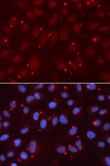
- Immunofluorescence analysis of XRCC3 in U2OS cells. Samples were incubated with XRCC3 polyclonal antibody (Product # PA5-76005).

- Submitted by
- Invitrogen Antibodies (provider)
- Main image
- Experimental details
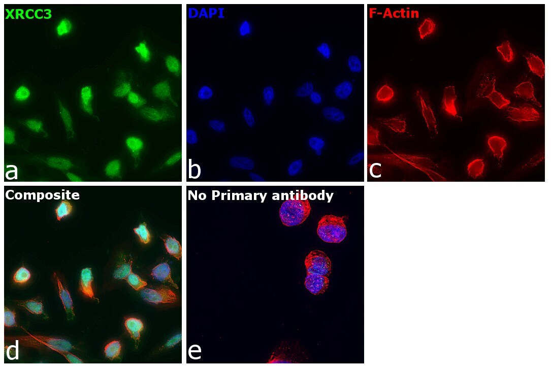
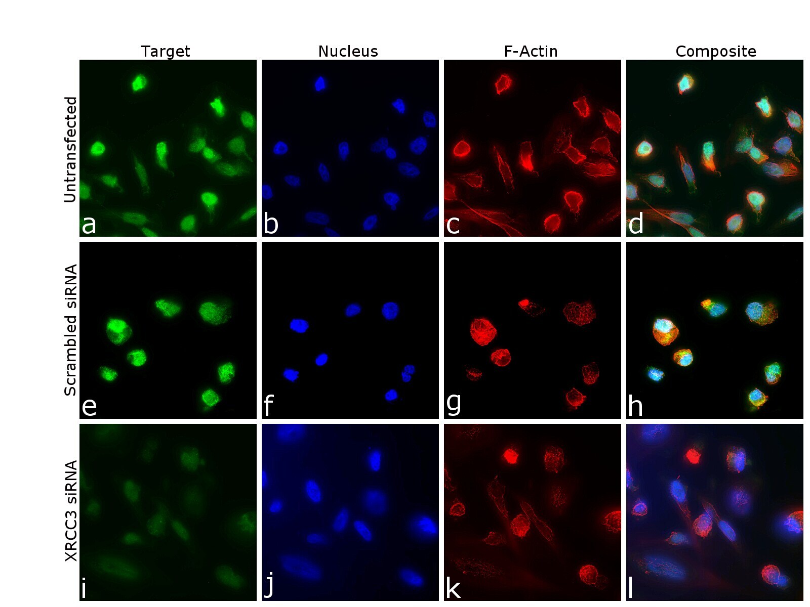
- Immunofluorescence analysis of DNA repair protein XRCC3 was performed using 70% confluent log phase PC-3 cells. The cells were fixed with 4% paraformaldehyde for 10 minutes, permeabilized with 0.1% Triton™ X-100 for 15 minutes, and blocked with 2% BSA for 45 minutes at room temperature. The cells were labeled with XRCC3 Polyclonal Antibody (Product # PA5-76005) at 1:100 dilution in 0.1% BSA, incubated at 4 degree celsius overnight and then labeled with Goat anti-Rabbit IgG (H+L) Highly Cross-Adsorbed Secondary Antibody, Alexa Fluor Plus 488 (Product # A32731), (1:2000 dilution), for 45 minutes at room temperature (Panel a: Green). Nuclei (Panel b:Blue) were stained with ProLong™ Diamond Antifade Mountant with DAPI (Product # P36962). F-actin (Panel c: Red) was stained with Rhodamine Phalloidin (Product # R415, 1:300 dilution). Panel d represents the merged image showing nucleus and cytoplasm localization. Panel e represents control cells with no primary antibody to assess background. The images were captured at 60X magnification in EVOS™ M7000 Imaging System (Product # AMF7000) and externally deconvoluted (D.Sage et al. / Methods 115 (2017) 28-41).

- Submitted by
- Invitrogen Antibodies (provider)
- Main image

- Experimental details
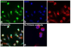
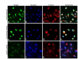
- Knockdown of DNA repair protein XRCC3 was achieved by transfecting PC-3 cells with DNA repair protein XRCC3 specific siRNA (Silencer® select Product # s14946, s14948). Immunofluorescence analysis was performed on untransfected PC-3 cells (panel a,d), transfected with non-specific scrambled siRNA (panels b,e) and transfected with DNA repair protein XRCC3 specific siRNA (panel c,f). Cells were fixed, permeabilized, and labelled with XRCC3 Polyclonal Antibody (Product # PA5-76005, 1:100 dilution) followed by Goat anti-Rabbit IgG (H+L) Highly Cross-Adsorbed Secondary Antibody, Alexa Fluor Plus 488 (Product # A32731), (1:2000 dilution). Nuclei (blue) were stained using ProLong™ Diamond Antifade Mountant with DAPI (Product # P36962), and Rhodamine Phalloidin (Product # R415, 1:300 dilution) was used for cytoskeletal F-actin (Red) staining. Reduction of specific signal was observed upon siRNA mediated knockdown (panel c,f) confirming specificity of the antibody to DNA repair protein XRCC3 (Green). The images were captured at 60X magnification in EVOS™ M7000 Imaging System (Product # AMF7000) and externally deconvoluted (D.Sage et al. / Methods 115 (2017) 28-41).

- Submitted by
- Invitrogen Antibodies (provider)
- Main image

- Experimental details
- Immunofluorescence analysis of XRCC3 in U2OS cells. Samples were incubated with XRCC3 polyclonal antibody (Product # PA5-76005).
- Submitted by
- Invitrogen Antibodies (provider)
- Main image

- Experimental details
- Immunofluorescence analysis of DNA repair protein XRCC3 was performed using 70% confluent log phase PC-3 cells. The cells were fixed with 4% paraformaldehyde for 10 minutes, permeabilized with 0.1% Triton™ X-100 for 15 minutes, and blocked with 2% BSA for 45 minutes at room temperature. The cells were labeled with XRCC3 Polyclonal Antibody (Product # PA5-76005) at 1:100 dilution in 0.1% BSA, incubated at 4 degree celsius overnight and then labeled with Goat anti-Rabbit IgG (H+L) Highly Cross-Adsorbed Secondary Antibody, Alexa Fluor Plus 488 (Product # A32731), (1:2000 dilution), for 45 minutes at room temperature (Panel a: Green). Nuclei (Panel b:Blue) were stained with ProLong™ Diamond Antifade Mountant with DAPI (Product # P36962). F-actin (Panel c: Red) was stained with Rhodamine Phalloidin (Product # R415, 1:300 dilution). Panel d represents the merged image showing nucleus and cytoplasm localization. Panel e represents control cells with no primary antibody to assess background. The images were captured at 60X magnification in EVOS™ M7000 Imaging System (Product # AMF7000) and externally deconvoluted (D.Sage et al. / Methods 115 (2017) 28-41).
- Submitted by
- Invitrogen Antibodies (provider)
- Main image
- Experimental details
- Knockdown of DNA repair protein XRCC3 was achieved by transfecting PC-3 cells with DNA repair protein XRCC3 specific siRNA (Silencer® select Product # s14946, s14948). Immunofluorescence analysis was performed on untransfected PC-3 cells (panel a,d), transfected with non-specific scrambled siRNA (panels b,e) and transfected with DNA repair protein XRCC3 specific siRNA (panel c,f). Cells were fixed, permeabilized, and labelled with XRCC3 Polyclonal Antibody (Product # PA5-76005, 1:100 dilution) followed by Goat anti-Rabbit IgG (H+L) Highly Cross-Adsorbed Secondary Antibody, Alexa Fluor Plus 488 (Product # A32731), (1:2000 dilution). Nuclei (blue) were stained using ProLong™ Diamond Antifade Mountant with DAPI (Product # P36962), and Rhodamine Phalloidin (Product # R415, 1:300 dilution) was used for cytoskeletal F-actin (Red) staining. Reduction of specific signal was observed upon siRNA mediated knockdown (panel c,f) confirming specificity of the antibody to DNA repair protein XRCC3 (Green). The images were captured at 60X magnification in EVOS™ M7000 Imaging System (Product # AMF7000) and externally deconvoluted (D.Sage et al. / Methods 115 (2017) 28-41).
Supportive validation
- Submitted by
- Invitrogen Antibodies (provider)
- Main image
- Experimental details
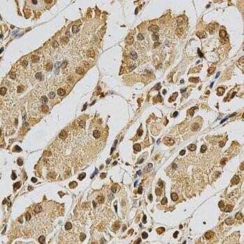
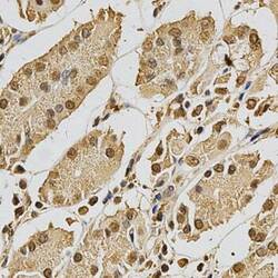
- Immunohistochemistry analysis of XRCC3 in paraffin-embedded human stomach tissue. Samples were incubated with XRCC3 polyclonal antibody (Product # PA5-76005).