Explore
 Validate
 Learn
Learn Western blot
Antibody data
- Antibody Data
- Antigen structure
- References [0]
- Comments [0]
- Validations
- Western blot [1]
- Immunocytochemistry [1]
- Immunohistochemistry [1]
- Flow cytometry [1]
Submit
Validation data
Reference
Comment
Report error
- Product number
- AMM85909 - Provider product page

- Provider
- EnkiLife Biotech Co., Ltd.
- Product name
- ITA6 Mouse Monoclonal Antibody
- Antibody type
- Monoclonal
- Description
- Affinity Purification
- Reactivity
- Human, Mouse
- Host
- Mouse
- Conjugate
- Unconjugated
- Antibody clone number
- Monoclonal
- Vial size
- 100 µl
- Concentration
- 1 mg/ml
- Storage
- Store at 4°C short term. Aliquot and store at -20°C long term. Avoid freeze/thaw cycles.
- Handling
- The antibody solution should be gently mixed before use.
No comments: Submit comment
Supportive validation
- Submitted by
- EnkiLife Biotech Co., Ltd. (provider)
- Main image

- Experimental details
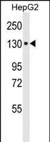
- Western blot analysis of ITA6 Mouse Monoclonal Antibody in HepG2 cell line lysates (35μg/lane). ITA6 (arrow) was detected using the purified Mab.(8μg/ml)
Supportive validation
- Submitted by
- EnkiLife Biotech Co., Ltd. (provider)
- Main image
- Experimental details
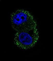
- Confocal immunofluorescent analysis of ITA6 Antibody (Cat#AMM85909) with HepG2 cell followed by Alexa Fluor® 488-conjugated goat anti-mouse lgG (green). DAPI was used to stain the cell nuclear (blue).
Supportive validation
- Submitted by
- EnkiLife Biotech Co., Ltd. (provider)
- Main image
- Experimental details
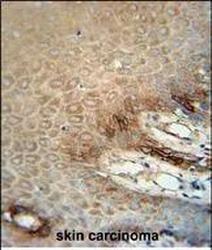
- ITA6 Monoclonal Antibody (Cat. #AMM85909) immunohistochemistry analysis in formalin fixed and paraffin embedded human skin carcinoma followed by peroxidase conjugation of the secondary antibody and DAB staining. This data demonstrates the use of the ITA6 Monoclonal Antibody for immunohistochemistry. Clinical relevance has not been evaluated.

Supportive validation
- Submitted by
- EnkiLife Biotech Co., Ltd. (provider)
- Main image
- Experimental details
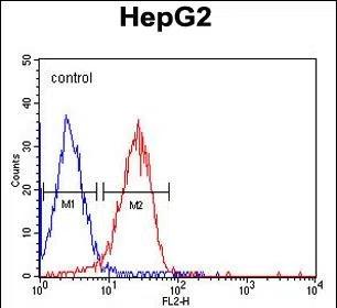
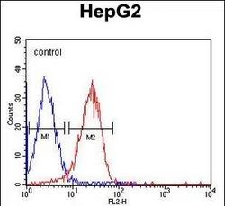
- ITA6 Monoclonal Antibody (Cat. #AMM85909) flow cytometric analysis of HepG2 cells (right histogram) compared to a negative control cell (left histogram).PE-conjugated goat-anti-mouse secondary antibodies were used for the analysis.