Explore
 Validate
 Learn
Learn Western blot
 Immunohistochemistry
Antibody data
- Antibody Data
- Antigen structure
- References [12]
- Comments [0]
- Validations
- Immunohistochemistry [1]
Submit
Validation data
Reference
Comment
Report error
- Product number
- HPA012696 - Provider product page

- Provider
- Atlas Antibodies
- Proper citation
- Atlas Antibodies Cat#HPA012696, RRID:AB_1851822
- Product name
- Anti-ITGA6
- Antibody type
- Polyclonal
- Description
- Polyclonal Antibody against Human ITGA6, Gene description: integrin, alpha 6, Alternative Gene Names: CD49f, Validated applications: WB, IHC, Uniprot ID: P23229, Storage: Store at +4°C for short term storage. Long time storage is recommended at -20°C.
- Reactivity
- Human
- Host
- Rabbit
- Conjugate
- Unconjugated
- Isotype
- IgG
- Vial size
- 100 µl
- Concentration
- 0.1 mg/ml
- Storage
- Store at +4°C for short term storage. Long time storage is recommended at -20°C.
- Handling
- The antibody solution should be gently mixed before use.
Submitted references Native Autoantigen Complex Detects Pemphigoid Autoantibodies
Epithelial cell attachment and adhesion protein expression on novel in sol TiO2 coated zirconia and titanium alloy surfaces
FAK inhibition alone or in combination with adjuvant therapies reduces cancer stem cell activity
Role of integrins in the metastatic spread of high-grade serous ovarian cancer
Dual Role of Integrin Alpha-6 in Glioblastoma: Supporting Stemness in Proneural Stem-Like Cells While Inducing Radioresistance in Mesenchymal Stem-Like Cells
BR-BCSC Signature: The Cancer Stem Cell Profile Enriched in Brain Metastases that Predicts a Worse Prognosis in Lymph Node-Positive Breast Cancer
Deep sequencing-based microRNA expression signatures in head and neck squamous cell carcinoma: dual strands of pre-miR-150 as antitumor miRNAs
P-cadherin: a useful biomarker for axillary-based breast cancer decisions in the clinical practice
Overexpression of Thy1 and ITGA6 is associated with invasion, metastasis and poor prognosis in human gallbladder carcinoma
Matrix Metalloproteinase 10 Degradomics in Keratinocytes and Epidermal Tissue Identifies Bioactive Substrates With Pleiotropic Functions*
Multiplex Flow Cytometry Barcoding and Antibody Arrays Identify Surface Antigen Profiles of Primary and Metastatic Colon Cancer Cell Lines
Identification of integrins α6 and β7 as c‐Jun‐ and transformation‐relevant genes in highly invasive fibrosarcoma cells
Mai S, Izumi K, Mai Y, Natsuga K, Ishii N, Sawamura D, Schauer F, Kiritsi D, Nishie W, Ujiie H
JID Innovations 2023;3(3):100193
JID Innovations 2023;3(3):100193
Epithelial cell attachment and adhesion protein expression on novel in sol TiO2 coated zirconia and titanium alloy surfaces
Riivari S, Närvä E, Kangasniemi I, Willberg J, Närhi T
Journal of Biomedical Materials Research Part B: Applied Biomaterials 2022;110(11):2533-2541
Journal of Biomedical Materials Research Part B: Applied Biomaterials 2022;110(11):2533-2541
FAK inhibition alone or in combination with adjuvant therapies reduces cancer stem cell activity
Timbrell S, Aglan H, Cramer A, Foden P, Weaver D, Pachter J, Kilgallon A, Clarke R, Farnie G, Bundred N
npj Breast Cancer 2021;7(1)
npj Breast Cancer 2021;7(1)
Role of integrins in the metastatic spread of high-grade serous ovarian cancer
Krajnak S, Jäkel J, Anić K, Schwab R, Schmidt M, Hasenburg A, Roth W, Brenner W, Battista M
Archives of Gynecology and Obstetrics 2021;305(5):1291-1298
Archives of Gynecology and Obstetrics 2021;305(5):1291-1298
Dual Role of Integrin Alpha-6 in Glioblastoma: Supporting Stemness in Proneural Stem-Like Cells While Inducing Radioresistance in Mesenchymal Stem-Like Cells
Stanzani E, Pedrosa L, Bourmeau G, Anezo O, Noguera-Castells A, Esteve-Codina A, Passoni L, Matteoli M, de la Iglesia N, Seano G, Martínez-Soler F, Tortosa A
Cancers 2021;13(12):3055
Cancers 2021;13(12):3055
BR-BCSC Signature: The Cancer Stem Cell Profile Enriched in Brain Metastases that Predicts a Worse Prognosis in Lymph Node-Positive Breast Cancer
Dionísio M, Vieira A, Carvalho R, Conde I, Oliveira M, Gomes M, Pinto M, Pereira P, Pimentel J, Souza C, Marques M, Duval da Silva V, Barroso A, Preto D, Cameselle-Teijeiro J, Schmitt F, Ribeiro A, Paredes J
Cells 2020;9(11):2442
Cells 2020;9(11):2442
Deep sequencing-based microRNA expression signatures in head and neck squamous cell carcinoma: dual strands of pre-miR-150 as antitumor miRNAs
Koshizuka K, Nohata N, Hanazawa T, Kikkawa N, Arai T, Okato A, Fukumoto I, Katada K, Okamoto Y, Seki N
Oncotarget 2017;8(18):30288-30304
Oncotarget 2017;8(18):30288-30304
P-cadherin: a useful biomarker for axillary-based breast cancer decisions in the clinical practice
Vieira A, Dionísio M, Gomes M, Cameselle-Teijeiro J, Lacerda M, Amendoeira I, Schmitt F, Paredes J
Modern Pathology 2017;30(5):698-709
Modern Pathology 2017;30(5):698-709
Overexpression of Thy1 and ITGA6 is associated with invasion, metastasis and poor prognosis in human gallbladder carcinoma
Zhang D, Yang Z, Zhou E, Miao X, Zou Q, Li J, Liang L, Zeng G, Chen S
Oncology Letters 2016;12(6):5136-5144
Oncology Letters 2016;12(6):5136-5144
Matrix Metalloproteinase 10 Degradomics in Keratinocytes and Epidermal Tissue Identifies Bioactive Substrates With Pleiotropic Functions*
Schlage P, Kockmann T, Sabino F, Kizhakkedathu J, auf dem Keller U
Molecular & Cellular Proteomics 2015;14(12):3234-3246
Molecular & Cellular Proteomics 2015;14(12):3234-3246
Multiplex Flow Cytometry Barcoding and Antibody Arrays Identify Surface Antigen Profiles of Primary and Metastatic Colon Cancer Cell Lines
Ulasov I, Sukhdeo K, Paramban R, Vidal J, Elia J, Martin J, Rivera M, Carrasco D, Jarrar A, Kalady M, Carson C, Balderas R, Hjelmeland A, Lathia J, Rich J
PLoS ONE 2013;8(1):e53015
PLoS ONE 2013;8(1):e53015
Identification of integrins α6 and β7 as c‐Jun‐ and transformation‐relevant genes in highly invasive fibrosarcoma cells
Kielosto M, Nummela P, Järvinen K, Yin M, Hölttä E
International Journal of Cancer 2009;125(5):1065-1073
International Journal of Cancer 2009;125(5):1065-1073
No comments: Submit comment
Supportive validation
- Submitted by
- Atlas Antibodies (provider)
- Enhanced method
- Orthogonal validation
- Main image
- Experimental details
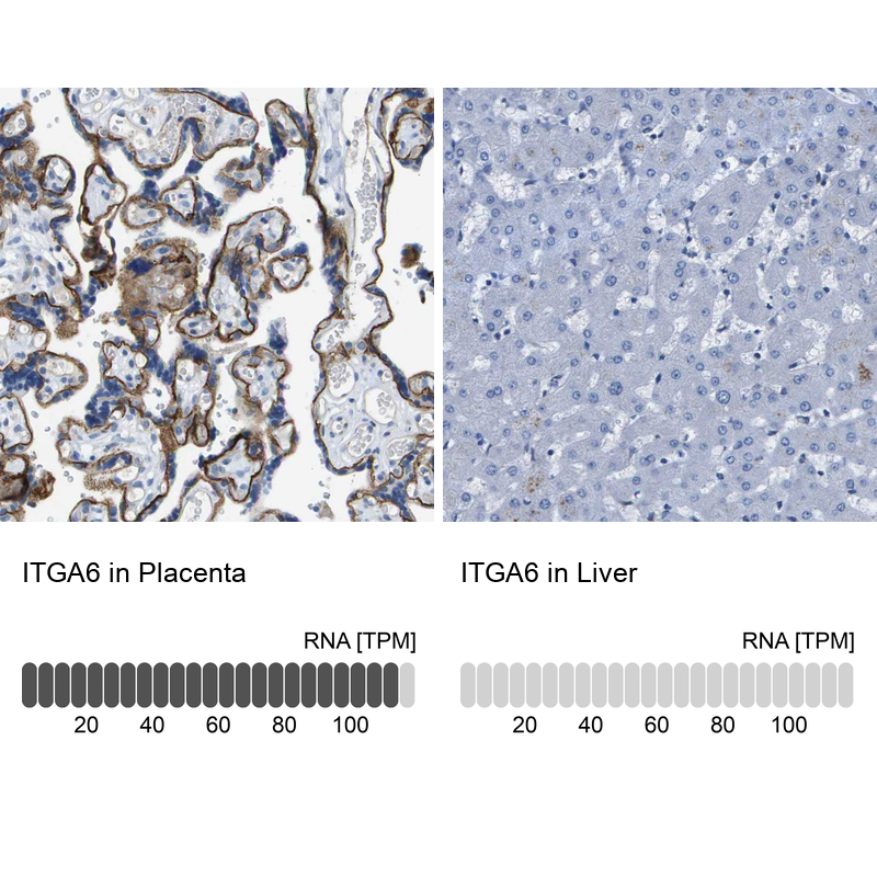
- Immunohistochemistry analysis in human placenta and liver tissues using HPA012696 antibody. Corresponding ITGA6 RNA-seq data are presented for the same tissues.
- Sample type
- Human
- Protocol
- Protocol