Explore
 Validate
 Learn
Learn Western blot
 ELISA
ELISAAntibody data
- Antibody Data
- Antigen structure
- References [0]
- Comments [0]
- Validations
- Western blot [2]
- Immunocytochemistry [3]
- Immunohistochemistry [8]
- Flow cytometry [2]
Submit
Validation data
Reference
Comment
Report error
- Product number
- RQ5770 - Provider product page

- Provider
- NSJ Bioreagents
- Product name
- TLN1 Antibody / Talin 1
- Antibody type
- Polyclonal
- Description
- This highly specific TLN1 antibody is suitable for use in Western blot/Immunohistochemistry/Immunofluorescence/Flow cytometry/Direct ELISA applications with human, mouse and rat samples.
- Reactivity
- Human, Mouse, Rat
- Host
- Rabbit
- Conjugate
- Unconjugated
- Vial size
- 100 ug
- Concentration
- 0.5mg/ml if reconstituted with 0.2ml sterile DI water
- Storage
- After reconstitution, the TLN1 antibody can be stored for up to one month at 4oC. For long-term, aliquot and store at -20oC. Avoid repeated freezing and thawing.
No comments: Submit comment
Supportive validation
- Submitted by
- NSJ Bioreagents (provider)
- Main image
- Experimental details
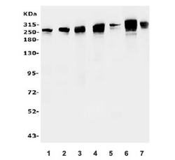
- Western blot testing of 1) human SW620, 2) human Raji, 3) human HEK293, 4) rat lung, 5) rat C6, 6) mouse lung and 7) mouse NIH 3T3 lysate with TLN1 antibody. Predicted molecular weight ~275 kDa.

- Submitted by
- NSJ Bioreagents (provider)
- Main image
- Experimental details
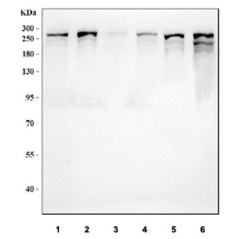
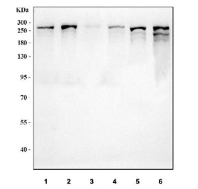
- Western blot testing of 1) human HeLa, 2) human HepG2, 3) human Raji, 4) human 293T, 5) rat C6 and 6) mouse lung tissue lysate with TLN1 antibody. Predicted molecular weight ~275 kDa.
Supportive validation
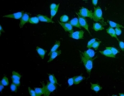
- Submitted by
- NSJ Bioreagents (provider)
- Main image

- Experimental details
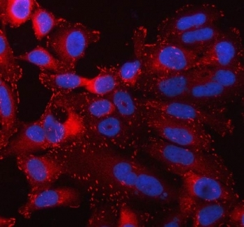
- Immunofluorescent staining of FFPE human HeLa cells with TLN1 antibody (green) and DAPI nuclear stain (blue). HIER: steam section in pH6 citrate buffer for 20 min.

- Submitted by
- NSJ Bioreagents (provider)
- Main image
- Experimental details
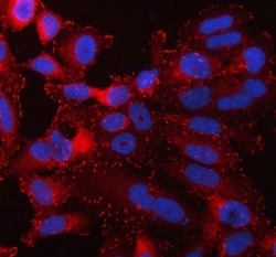
- Immunofluorescent staining of FFPE human HeLa cells with TLN1 antibody (red) and DAPI nuclear stain (blue). HIER: steam section in pH6 citrate buffer for 20 min.
- Submitted by
- NSJ Bioreagents (provider)
- Main image
- Experimental details
- Immunofluorescent staining of FFPE human HeLa cells with TLN1 antibody (green) and DAPI nuclear stain (blue). HIER: steam section in pH6 citrate buffer for 20 min.

Supportive validation
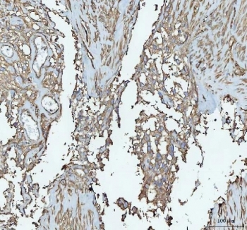
- Submitted by
- NSJ Bioreagents (provider)
- Main image

- Experimental details
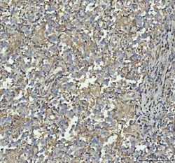
- IHC staining of FFPE human renal cancer with TLN1 antibody. HIER: boil tissue sections in pH8 EDTA for 20 min and allow to cool before testing.

- Submitted by
- NSJ Bioreagents (provider)
- Main image

- Experimental details
- IHC staining of FFPE human bladder urothelial carcinoma tissue with TLN1 antibody. HIER: boil tissue sections in pH8 EDTA for 20 min and allow to cool before testing.
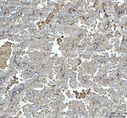
- Submitted by
- NSJ Bioreagents (provider)
- Main image

- Experimental details
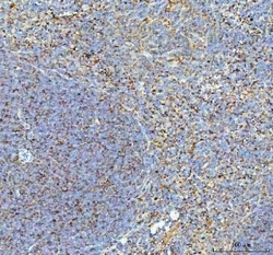
- IHC staining of FFPE human tonsil tissue with TLN1 antibody. HIER: boil tissue sections in pH8 EDTA for 20 min and allow to cool before testing.

- Submitted by
- NSJ Bioreagents (provider)
- Main image
- Experimental details
- IHC staining of FFPE human testicular germ cell tumor tissue with TLN1 antibody. HIER: boil tissue sections in pH8 EDTA for 20 min and allow to cool before testing.

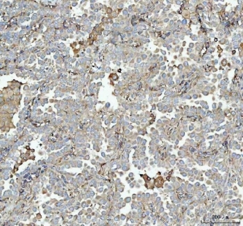
- Submitted by
- NSJ Bioreagents (provider)
- Main image
- Experimental details
- IHC staining of FFPE human lung cancer tissue with TLN1 antibody. HIER: boil tissue sections in pH8 EDTA for 20 min and allow to cool before testing.
- Submitted by
- NSJ Bioreagents (provider)
- Main image
- Experimental details
- IHC staining of FFPE human renal cancer tissue with TLN1 antibody. HIER: boil tissue sections in pH8 EDTA for 20 min and allow to cool before testing.

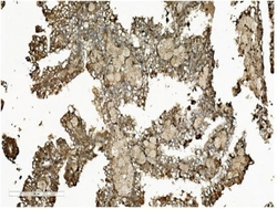
- Submitted by
- NSJ Bioreagents (provider)
- Main image
- Experimental details
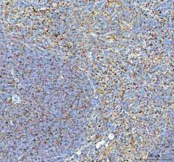
- IHC staining of FFPE mouse spleen tissue with TLN1 antibody. HIER: boil tissue sections in pH8 EDTA for 20 min and allow to cool before testing.
- Submitted by
- NSJ Bioreagents (provider)
- Main image
- Experimental details
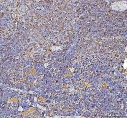
- IHC staining of FFPE rat spleen tissue with TLN1 antibody. HIER: boil tissue sections in pH8 EDTA for 20 min and allow to cool before testing.

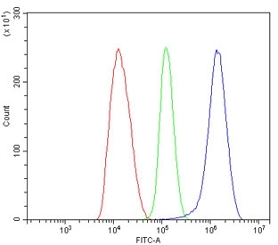
Supportive validation
- Submitted by
- NSJ Bioreagents (provider)
- Main image
- Experimental details
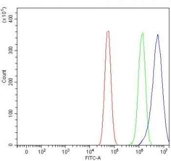
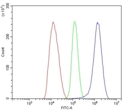
- Flow cytometry testing of human HepG2 cells with TLN1 antibody at 1ug/million cells (blocked with goat sera); Red=cells alone, Green=isotype control, Blue= TLN1 antibody.
- Submitted by
- NSJ Bioreagents (provider)
- Main image
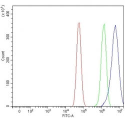
- Experimental details
- Flow cytometry testing of human U-87 MG cells with TLN1 antibody at 1ug/million cells (blocked with goat sera); Red=cells alone, Green=isotype control, Blue= TLN1 antibody.