Explore
 Validate
 Learn
Learn Western blot
 Immunocytochemistry
Antibody data
- Antibody Data
- Antigen structure
- References [2]
- Comments [0]
- Validations
- Immunocytochemistry [1]
Submit
Validation data
Reference
Comment
Report error
- Product number
- HPA004748 - Provider product page

- Provider
- Atlas Antibodies
- Proper citation
- Atlas Antibodies Cat#HPA004748, RRID:AB_1857760
- Product name
- Anti-TLN1
- Antibody type
- Polyclonal
- Description
- Polyclonal Antibody against Human TLN1, Gene description: talin 1, Alternative Gene Names: ILWEQ, TLN, Validated applications: ICC, IHC, WB, Uniprot ID: Q9Y490, Storage: Store at +4°C for short term storage. Long time storage is recommended at -20°C.
- Reactivity
- Human
- Host
- Rabbit
- Conjugate
- Unconjugated
- Isotype
- IgG
- Vial size
- 100 µl
- Concentration
- 0.1 mg/ml
- Storage
- Store at +4°C for short term storage. Long time storage is recommended at -20°C.
- Handling
- The antibody solution should be gently mixed before use.
Submitted references Phosphatidylinositol Phosphate 5-Kinase Iγi2 in Association with Src Controls Anchorage-independent Growth of Tumor Cells
Discs Large 1 (Dlg1) Scaffolding Protein Participates with Clathrin and Adaptator Protein Complex 1 (AP-1) in Forming Weibel-Palade Bodies of Endothelial Cells
Thapa N, Choi S, Hedman A, Tan X, Anderson R
Journal of Biological Chemistry 2013;288(48):34707-34718
Journal of Biological Chemistry 2013;288(48):34707-34718
Discs Large 1 (Dlg1) Scaffolding Protein Participates with Clathrin and Adaptator Protein Complex 1 (AP-1) in Forming Weibel-Palade Bodies of Endothelial Cells
Philippe M, Léger T, Desvaux R, Walch L
Journal of Biological Chemistry 2013;288(18):13046-13056
Journal of Biological Chemistry 2013;288(18):13046-13056
No comments: Submit comment
Supportive validation
- Submitted by
- Atlas Antibodies (provider)
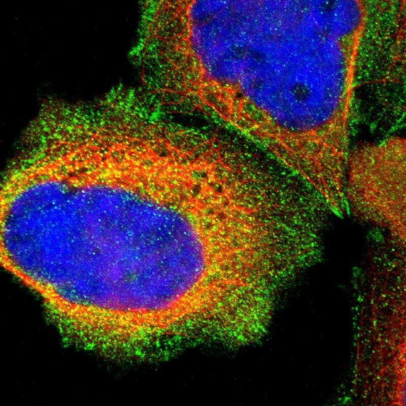
- Main image

- Experimental details
- Immunofluorescent staining of human cell line A-431 shows localization to plasma membrane, cytosol & focal adhesion sites.
- Sample type
- Human