Explore
 Validate
 Learn
Learn Western blot
 Flow cytometry
Antibody data
- Antibody Data
- Antigen structure
- References [1]
- Comments [0]
- Validations
- Western blot [4]
- Immunocytochemistry [2]
Submit
Validation data
Reference
Comment
Report error
- Product number
- PA5-47096 - Provider product page

- Provider
- Invitrogen Antibodies
- Product name
- CD51 Polyclonal Antibody
- Antibody type
- Polyclonal
- Antigen
- Recombinant full-length protein
- Description
- In Western blots, less than 1% cross-reactivity with recombinant human Integrin alpha 5 and recombinant mouse Integrin E is observed. Reconstitute at 0.2 mg/mL in sterile PBS.
- Reactivity
- Human, Mouse, Rat
- Host
- Goat
- Isotype
- IgG
- Vial size
- 100 µg
- Concentration
- 0.2 mg/mL
- Storage
- -20° C, Avoid Freeze/Thaw Cycles
Submitted references Wnt signaling regulates trans-differentiation of stem cell like type 2 alveolar epithelial cells to type 1 epithelial cells.
Abdelwahab EMM, Rapp J, Feller D, Csongei V, Pal S, Bartis D, Thickett DR, Pongracz JE
Respiratory research 2019 Sep 6;20(1):204
Respiratory research 2019 Sep 6;20(1):204
No comments: Submit comment
Supportive validation
- Submitted by
- Invitrogen Antibodies (provider)
- Main image
- Experimental details
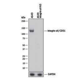
- Knockout validation by Western blot analysis of Integrin alpha V (CD51) in lysates of A549 human lung carcinoma parental cell line and Integrin αV/CD51 knockout A549 cell line (KO). Samples were incubated in Integrin alpha V (CD51) polyclonal antibody (Product # PA5-47096) using a dilution of 0.5 µg/mL followed by a HRP-conjugated Anti-Goat IgG secondary antibody. A specific band was detected for Integrin αV/CD51 at approximately 120 kDa (as indicated) in the parental A549 cell line, but is not detectable in knockout A549 cell line. GAPDH is shown as a loading control. This experiment was conducted under reducing conditions.

- Submitted by
- Invitrogen Antibodies (provider)
- Main image
- Experimental details
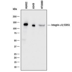
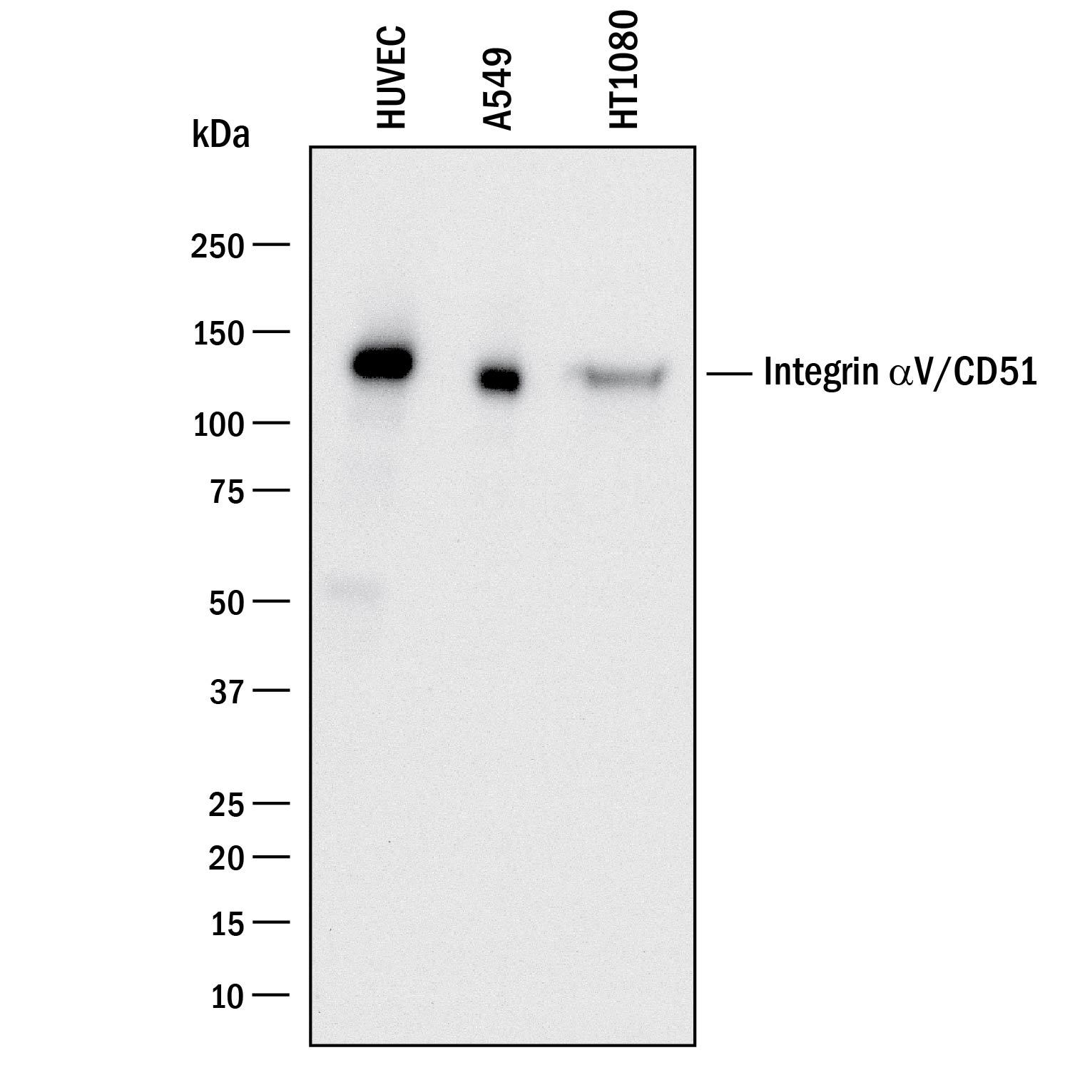
- Western blot analysis of CD51 in lysates of HUVEC human umbilical vein endothelial cells, A549 human lung carcinoma cell line, and HT1080 human fibrosarcoma cell line. Samples were incubated in CD51 polyclonal antibody (Product # PA5-47096) using a dilution of 0.5 µg/mL followed by a HRP-conjugated Anti-Goat IgG secondary antibody. A specific band was detected for Integrin αV/CD51 at approximately 130 kDa (as indicated). This experiment was conducted under reducing conditions.
- Submitted by
- Invitrogen Antibodies (provider)
- Main image
- Experimental details
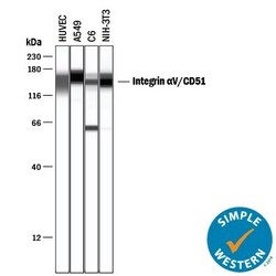
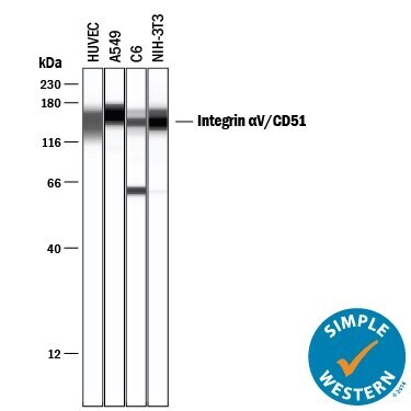
- Western blot analysis of Integrin alpha V (CD51) in 0.2 mg/mL lysates of HUVEC human umbilical vein endothelial cells, A549 human lung carcinoma cell line, C6 rat glioma cell line, and NIH‚3T3 mouse embryonic fibroblast cell line. Samples were incubated in Integrin alpha V (CD51) polyclonal antibody (Product # PA5-47096) using a dilution of 20 µg/mL followed by HRP-conjugated Anti-Goat IgG at a dilution of 0.0763888888888889. A specific band was detected for Integrin αV/CD51 at approximately 149-161 kDa (as indicated). This experiment was conducted under reducing conditions and using the 12-230 kDa separation system.
- Submitted by
- Invitrogen Antibodies (provider)
- Main image
- Experimental details
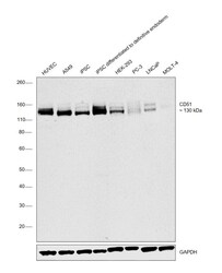
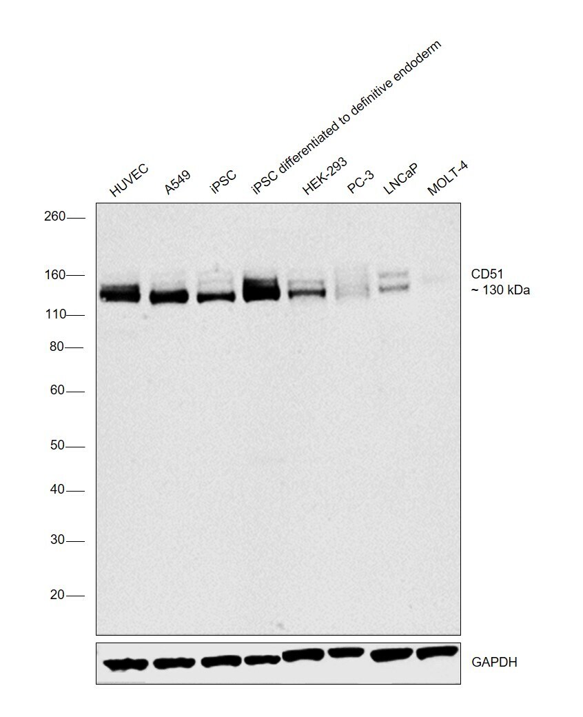
- Western blot was performed using Anti-CD51 Polyclonal Antibody (Product # PA5-47096) and a 130 kDa band corresponding to CD51 was observed across all the cell lines tested and the expression was increased in iPSC upon differentiation to definitive endoderm. Membrane enriched extracts (30 µg lysate) of HUVEC (Lane 1), A549 (Lane 2), iPSC (Lane 3), iPSC differentiated to definitive endoderm (Lane 4), HEK-293 (Lane 5), PC-3 (Lane 6), LNCaP (Lane 7) and MOLT-4 (Lane 8) were electrophoresed using Novex® NuPAGE® 4-12% Bis-Tris Protein Gel (Product # NP0322BOX). Resolved proteins were then transferred onto a nitrocellulose membrane (Product # IB23001) by iBlot® 2 Dry Blotting System (Product # IB21001). The blot was probed with the primary antibody (0.5ug/ml) and detected by chemiluminescence with Rabbit anti-Goat IgG (H+L), Superclonal™ Recombinant Secondary Antibody, HRP (Product # A27014, 1:4000 dilution) using the iBright FL 1000 (Product # A32752). Chemiluminescent detection was performed using Novex® ECL Chemiluminescent Substrate Reagent Kit (Product # WP20005).
Supportive validation
- Submitted by
- Invitrogen Antibodies (provider)
- Main image
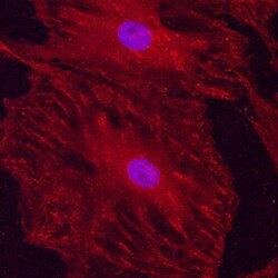
- Experimental details
- Immunocytochemistry analysis of CD51 in immersion fixed undifferentiated rat mesenchymal stem cells. Samples were incubated in CD51 polyclonal antibody (Product # PA5-47096) using a dilution of 10 µg/mL for 3 hours at room temperature followed by NorthernLights™ 557-conjugated Anti-Goat IgG Secondary Antibody (red) and counterstained with DAPI (blue). Specific staining was localized to cytoplasm.

- Submitted by
- Invitrogen Antibodies (provider)
- Main image
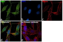
- Experimental details
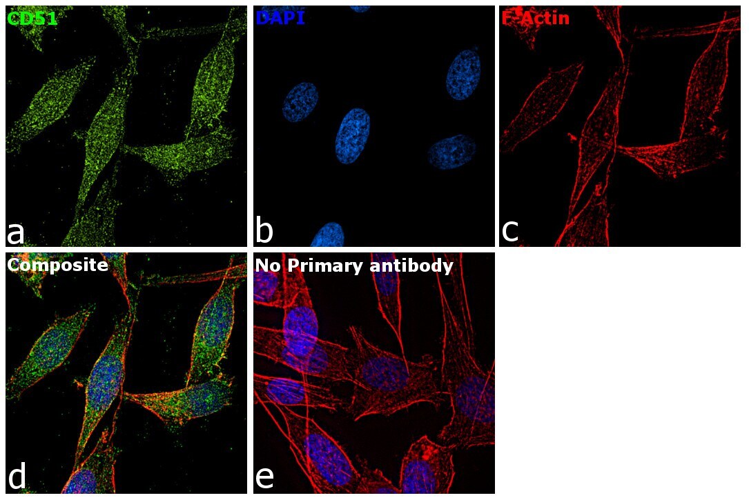
- Immunofluorescence analysis of CD51 was performed using 70% confluent log phase PC-3 cells. The cells were fixed with 4% paraformaldehyde for 10 minutes, permeabilized with 0.1% Triton™ X-100 for 15 minutes, and blocked with 2% BSA for 1 hour at room temperature. The cells were labeled with CD51 Polyclonal Antibody (Product # PA5-47096) at 5 µg/mL in 0.1% BSA, incubated at 4 degree Celsius overnight and then labeled with Rabbit anti-Goat IgG (H+L) Superclonal™ Secondary Antibody, Alexa Fluor® 488 conjugate (Product # A-11078) at a dilution of 1:2000 for 45 minutes at room temperature (Panel a: green). Nuclei (Panel b: blue) were stained with ProLong™ Diamond Antifade Mountant with DAPI (Product # P36962). F-actin (Panel c: red) was stained with Rhodamine Phalloidin (Product # R415). Panel d represents the merged image showing Nuclear localization. Panel e represents control cells with no primary antibody to assess background. The images were captured at 60X magnification.