Explore
 Validate
 Learn
Learn Western blot
 ELISA
ELISAAntibody data
- Antibody Data
- Antigen structure
- References [3]
- Comments [0]
- Validations
- Western blot [1]
Submit
Validation data
Reference
Comment
Report error
- Product number
- A01561-2 - Provider product page

- Provider
- Boster Biological Technology
- Product name
- Anti-Integrin alpha V/ITGAV Antibody Picoband™
- Antibody type
- Polyclonal
- Description
- Rabbit IgG polyclonal antibody for Integrin alpha V/ITGAV detection. Tested with WB, FCM, Direct ELISA in Human.
- Reactivity
- Human
- Host
- Rabbit
- Vial size
- 100μg/vial
- Concentration
- Add 0.2ml of distilled water will yield a concentration of 500ug/ml.
- Storage
- At -20°C for one year. After reconstitution, at 4°C for one month. It can also be aliquoted and stored frozen at -20°C for a longer time. Avoid repeated freezing and thawing.
- Handling
- Add 0.2ml of distilled water will yield a concentration of 500ug/ml.
Submitted references TGFβ3-mediated induction of Periostin facilitates head and neck cancer growth and is associated with metastasis.
Study on the expression and clinical significances of lewis y antigen and integrin αv, β3 in epithelial ovarian tumors.
Overexpression of Lewis(y) antigen protects ovarian cancer RMG-1 cells from carboplatin-induced apoptosis by the upregulation of Topo-I and Topo-II β.
Qin X, Yan M, Zhang J, Wang X, Shen Z, Lv Z, Li Z, Wei W, Chen W
Scientific reports 2016 Feb 9;6:20587
Scientific reports 2016 Feb 9;6:20587
Study on the expression and clinical significances of lewis y antigen and integrin αv, β3 in epithelial ovarian tumors.
Wang Y, Liu J, Lin B, Wang C, Li Q, Liu S, Yan L, Zhang S, Iwamori M
International journal of molecular sciences 2011;12(6):3409-21
International journal of molecular sciences 2011;12(6):3409-21
Overexpression of Lewis(y) antigen protects ovarian cancer RMG-1 cells from carboplatin-induced apoptosis by the upregulation of Topo-I and Topo-II β.
Wang C, Yan L, Wang Y, Lin B, Liu S, Li Q, Gao L, Zhang S, Iwamori M
Anatomical record (Hoboken, N.J. : 2007) 2011 Jun;294(6):961-9
Anatomical record (Hoboken, N.J. : 2007) 2011 Jun;294(6):961-9
No comments: Submit comment
Supportive validation
- Submitted by
- Boster Biological Technology (provider)
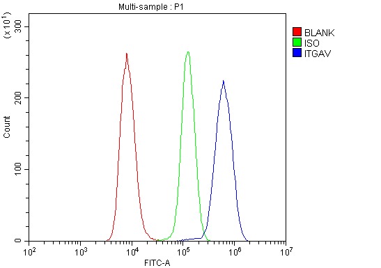
- Main image
- Experimental details
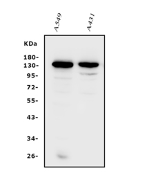
- Western blot analysis of Integrin alpha V/ITGAV using anti-Integrin alpha V/ITGAV antibody (A01561-2). Electrophoresis was performed on a 5-20% SDS-PAGE gel at 70V (Stacking gel) / 90V (Resolving gel) for 2-3 hours. The sample well of each lane was loaded with 50ug of sample under reducing conditions. Lane 1: human A549 whole cell lysates, Lane 2: human A431 whole cell lysates. After Electrophoresis, proteins were transferred to a Nitrocellulose membrane at 150mA for 50-90 minutes. Blocked the membrane with 5% Non-fat Milk/ TBS for 1.5 hour at RT. The membrane was incubated with rabbit anti-Integrin alpha V/ITGAV antigen affinity purified polyclonal antibody (Catalog # A01561-2) at 0.25 μg/mL overnight at 4°C, then washed with TBS-0.1%Tween 3 times with 5 minutes each and probed with a goat anti-rabbit IgG-HRP secondary antibody at a dilution of 1:10000 for 1.5 hour at RT. The signal is developed using an Enhanced Chemiluminescent detection (ECL) kit (Catalog # EK1002) with Tanon 5200 system. A specific band was detected for Integrin alpha V/ITGAV at approximately 130-140KD. The expected band size for Integrin alpha V/ITGAV is at 130KD.
- Additional image