Explore
 Validate
 Learn
Learn Western blot
 Flow cytometry
Antibody data
- Antibody Data
- Antigen structure
- References [3]
- Comments [0]
- Validations
- Western blot [2]
- Immunocytochemistry [1]
Submit
Validation data
Reference
Comment
Report error
- Product number
- AF1219 - Provider product page

- Provider
- R&D Systems
- Product name
- Human/Mouse/Rat Integrin alpha V/CD51 Antibody
- Antibody type
- Polyclonal
- Description
- Antigen Affinity-purified. Detects human Integrin alpha V in direct ELISAs and Western blots. Detects human, mouse and rat Integrin alpha V in Simple Western application. In Western blots, less than 1% cross-reactivity with recombinant human Integrin alpha 5 and recombinant mouse Integrin E is observed.
- Reactivity
- Human, Mouse, Rat
- Host
- Goat
- Conjugate
- Unconjugated
- Antigen sequence
P06756- Isotype
- IgG
- Vial size
- 100 ug
- Concentration
- LYOPH
- Storage
- Use a manual defrost freezer and avoid repeated freeze-thaw cycles. 12 months from date of receipt, -20 to -70 °C as supplied. 1 month, 2 to 8 °C under sterile conditions after reconstitution. 6 months, -20 to -70 °C under sterile conditions after reconstitution.
Submitted references Characterization of Human and Murine T-Cell Immunoglobulin Mucin Domain 4 (TIM-4) IgV Domain Residues Critical for Ebola Virus Entry.
Quantity and accessibility for specific targeting of receptors in tumours.
Binding of extracellular maspin to beta1 integrins inhibits vascular smooth muscle cell migration.
Rhein BA, Brouillette RB, Schaack GA, Chiorini JA, Maury W
Journal of virology 2016 Jul 1;90(13):6097-6111
Journal of virology 2016 Jul 1;90(13):6097-6111
Quantity and accessibility for specific targeting of receptors in tumours.
Hussain S, Rodriguez-Fernandez M, Braun GB, Doyle FJ 3rd, Ruoslahti E
Scientific reports 2014 Jun 10;4:5232
Scientific reports 2014 Jun 10;4:5232
Binding of extracellular maspin to beta1 integrins inhibits vascular smooth muscle cell migration.
Bass R, Wagstaff L, Ravenhill L, Ellis V
The Journal of biological chemistry 2009 Oct 2;284(40):27712-20
The Journal of biological chemistry 2009 Oct 2;284(40):27712-20
No comments: Submit comment
Supportive validation
- Submitted by
- R&D Systems (provider)
- Main image

- Experimental details
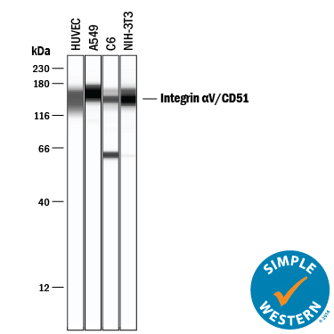
- Detection of Human, Mouse, and Rat Integrin alpha V/CD51 by Simple WesternTM. Simple Western lane view shows lysates of HUVEC human umbilical vein endothelial cells, A549 human lung carcinoma cell line, C6 rat glioma cell line, and NIH-3T3 mouse embryonic fibroblast cell line, loaded at 0.2 mg/mL. A specific band was detected for Integrin alpha V/CD51 at approximately 149-161 kDa (as indicated) using 20 µg/mL of Goat Anti-Human/Mouse/Rat Integrin alpha V/CD51 Antigen Affinity-purified Polyclonal Antibody (Catalog # AF1219) followed by 1:50 dilution of HRP-conjugated Anti-Goat IgG Secondary Antibody (Catalog # HAF109). This experiment was conducted under reducing conditions and using the 12-230 kDa separation system.
- Submitted by
- R&D Systems (provider)
- Main image
- Experimental details
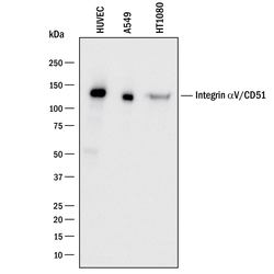
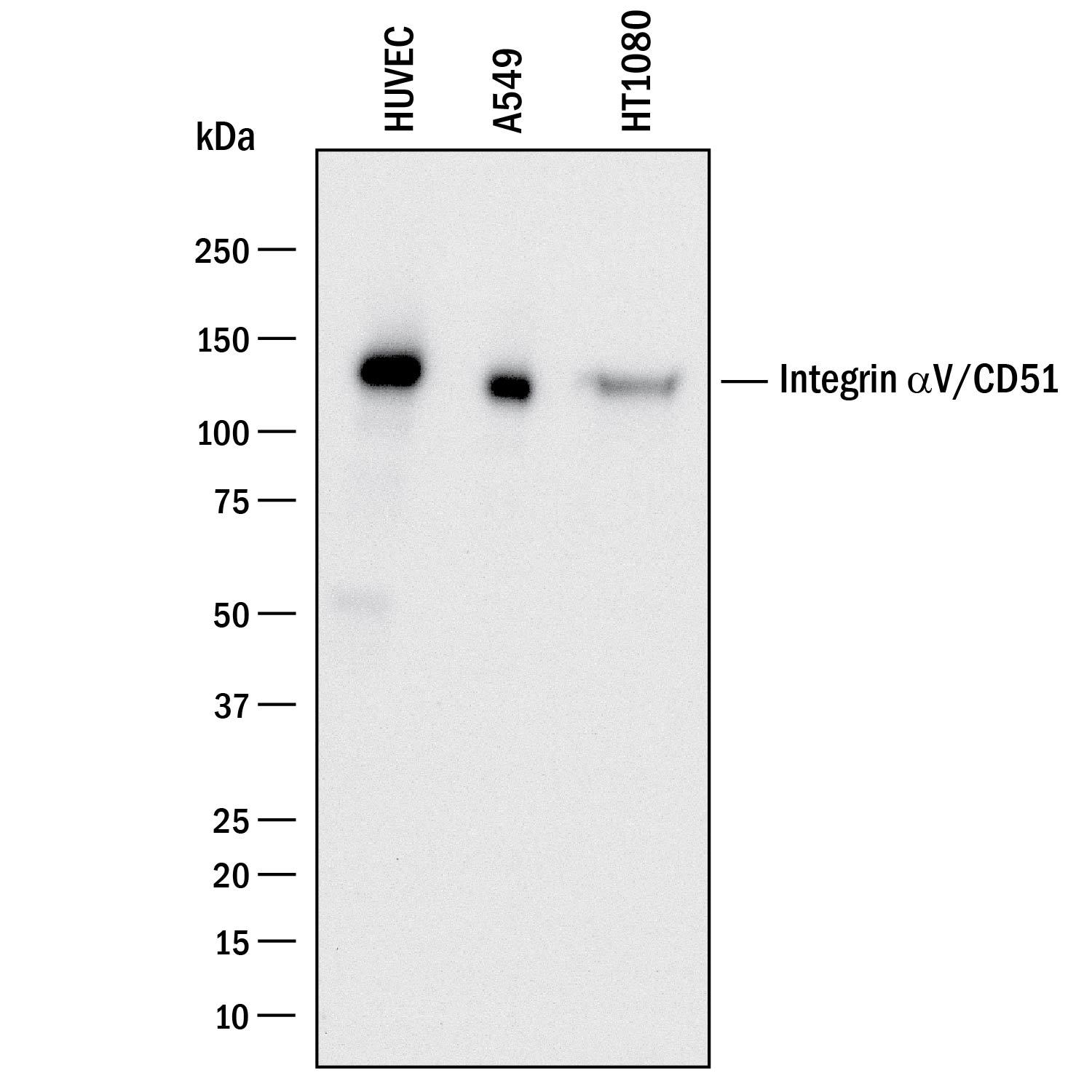
- Detection of Human Integrin alpha V/CD51 by Western Blot. Western blot shows lysates of HUVEC human umbilical vein endothelial cells, A549 human lung carcinoma cell line, and HT1080 human fibrosarcoma cell line. PVDF membrane was probed with 0.5 µg/mL of Goat Anti-Human Integrin alpha V/CD51 Antigen Affinity-purified Polyclonal Antibody (Catalog # AF1219) followed by HRP-conjugated Anti-Goat IgG Secondary Antibody (Catalog # HAF017). A specific band was detected for Integrin alpha V/CD51 at approximately 130 kDa (as indicated). This experiment was conducted under reducing conditions and using Immunoblot Buffer Group 1.
Supportive validation
- Submitted by
- R&D Systems (provider)
- Main image

- Experimental details
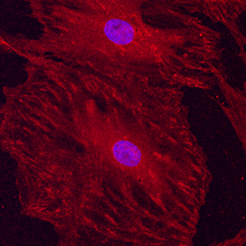
- Integrin alpha V/CD51 in Rat Mesenchymal Stem Cells. Integrin alpha V/CD51 was detected in immersion fixed undifferentiated rat mesenchymal stem cells using Goat Anti-Human/Mouse/Rat Integrin alpha V/CD51 Antigen Affinity-purified Polyclonal Antibody (Catalog # AF1219) at 10 µg/mL for 3 hours at room temperature. Cells were stained using the NorthernLights™ 557-conjugated Anti-Goat IgG Secondary Antibody (red; Catalog # NL001) and counterstained with DAPI (blue). Specific staining was localized to cytoplasm. View our protocol for Fluorescent ICC Staining of Cells on Coverslips.