Explore
 Validate
 Learn
Learn Western blot
 Immunoprecipitation
Antibody data
- Antibody Data
- Antigen structure
- References [11]
- Comments [0]
- Validations
- Western blot [1]
- Flow cytometry [1]
Submit
Validation data
Reference
Comment
Report error
- Product number
- 42-6200 - Provider product page

- Provider
- Invitrogen Antibodies
- Product name
- Anti-NDRG1 Polyclonal Antibody
- Antibody type
- Polyclonal
- Antigen
- Synthetic peptide
- Reactivity
- Human, Mouse
- Host
- Rabbit
- Isotype
- IgG
- Vial size
- 100 µg
- Concentration
- 0.25 mg/mL
- Storage
- -20°C
Submitted references Mucosa-Associated Lymphoid Tissue 1 Is an Oncogene Inducing Cell Proliferation, Invasion, and Tumor Growth via the Upregulation of NF-κB Activity in Human Prostate Carcinoma Cells.
Prostate-derived ets factor represses tumorigenesis and modulates epithelial-to-mesenchymal transition in bladder carcinoma cells.
The Iron Chelator, Dp44mT, Effectively Inhibits Human Oral Squamous Cell Carcinoma Cell Growth in Vitro and in Vivo.
N-myc downstream-regulated gene 1 (NDRG1) mediates pomegranate juice protection from apoptosis in hypoxic BeWo cells but not in primary human trophoblasts.
Growth differentiation factor-15: a p53- and demethylation-upregulating gene represses cell proliferation, invasion, and tumorigenesis in bladder carcinoma cells.
N-myc downstream-regulated gene 1 downregulates cell proliferation, invasiveness, and tumorigenesis in human oral squamous cell carcinoma.
Mechanisms by which interleukin-6 attenuates cell invasion and tumorigenesis in human bladder carcinoma cells.
Metallothionein 3: an androgen-upregulated gene enhances cell invasion and tumorigenesis of prostate carcinoma cells.
Glycoprotein transmembrane nmb: an androgen-downregulated gene attenuates cell invasion and tumorigenesis in prostate carcinoma cells.
Upregulation of prostate-derived Ets factor by luteolin causes inhibition of cell proliferation and cell invasion in prostate carcinoma cells.
L-Mimosine blocks cell proliferation via upregulation of B-cell translocation gene 2 and N-myc downstream regulated gene 1 in prostate carcinoma cells.
Tsui KH, Chang KS, Sung HC, Hsu SY, Lin YH, Hou CP, Yang PS, Chen CL, Feng TH, Juang HH
Biomedicines 2021 Mar 3;9(3)
Biomedicines 2021 Mar 3;9(3)
Prostate-derived ets factor represses tumorigenesis and modulates epithelial-to-mesenchymal transition in bladder carcinoma cells.
Tsui KH, Lin YH, Chung LC, Chuang ST, Feng TH, Chiang KC, Chang PL, Yeh CJ, Juang HH
Cancer letters 2016 May 28;375(1):142-151
Cancer letters 2016 May 28;375(1):142-151
The Iron Chelator, Dp44mT, Effectively Inhibits Human Oral Squamous Cell Carcinoma Cell Growth in Vitro and in Vivo.
Lee JC, Chiang KC, Feng TH, Chen YJ, Chuang ST, Tsui KH, Chung LC, Juang HH
International journal of molecular sciences 2016 Aug 31;17(9)
International journal of molecular sciences 2016 Aug 31;17(9)
N-myc downstream-regulated gene 1 (NDRG1) mediates pomegranate juice protection from apoptosis in hypoxic BeWo cells but not in primary human trophoblasts.
Chen B, Zaveri PG, Longtine MS, Nelson DM
Placenta 2015 Aug;36(8):847-53
Placenta 2015 Aug;36(8):847-53
Growth differentiation factor-15: a p53- and demethylation-upregulating gene represses cell proliferation, invasion, and tumorigenesis in bladder carcinoma cells.
Tsui KH, Hsu SY, Chung LC, Lin YH, Feng TH, Lee TY, Chang PL, Juang HH
Scientific reports 2015 Aug 7;5:12870
Scientific reports 2015 Aug 7;5:12870
N-myc downstream-regulated gene 1 downregulates cell proliferation, invasiveness, and tumorigenesis in human oral squamous cell carcinoma.
Lee JC, Chung LC, Chen YJ, Feng TH, Juang HH
Cancer letters 2014 Dec 28;355(2):242-52
Cancer letters 2014 Dec 28;355(2):242-52
Mechanisms by which interleukin-6 attenuates cell invasion and tumorigenesis in human bladder carcinoma cells.
Tsui KH, Wang SW, Chung LC, Feng TH, Lee TY, Chang PL, Juang HH
BioMed research international 2013;2013:791212
BioMed research international 2013;2013:791212
Metallothionein 3: an androgen-upregulated gene enhances cell invasion and tumorigenesis of prostate carcinoma cells.
Juang HH, Chung LC, Sung HC, Feng TH, Lee YH, Chang PL, Tsui KH
The Prostate 2013 Oct;73(14):1495-506
The Prostate 2013 Oct;73(14):1495-506
Glycoprotein transmembrane nmb: an androgen-downregulated gene attenuates cell invasion and tumorigenesis in prostate carcinoma cells.
Tsui KH, Chang YL, Feng TH, Chang PL, Juang HH
The Prostate 2012 Sep 15;72(13):1431-42
The Prostate 2012 Sep 15;72(13):1431-42
Upregulation of prostate-derived Ets factor by luteolin causes inhibition of cell proliferation and cell invasion in prostate carcinoma cells.
Tsui KH, Chung LC, Feng TH, Chang PL, Juang HH
International journal of cancer 2012 Jun 15;130(12):2812-23
International journal of cancer 2012 Jun 15;130(12):2812-23
L-Mimosine blocks cell proliferation via upregulation of B-cell translocation gene 2 and N-myc downstream regulated gene 1 in prostate carcinoma cells.
Chung LC, Tsui KH, Feng TH, Lee SL, Chang PL, Juang HH
American journal of physiology. Cell physiology 2012 Feb 15;302(4):C676-85
American journal of physiology. Cell physiology 2012 Feb 15;302(4):C676-85
No comments: Submit comment
Supportive validation
- Submitted by
- Invitrogen Antibodies (provider)
- Main image
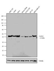
- Experimental details
- Western blot analysis was performed on whole cell extracts (30 µg lysate) of HEK 293 (Lane 1), Jurkat (Lane 2), HeLa (Lane 3), Mouse Brain (tissue extract) (Lane 4), COLO 205 (Lane 5), HT-29 (Lane 6) and MDA-MB-231 (Lane 7). The blot was probed with Anti-CAP43 Rabbit Polyclonal Antibody (Product # 42-6200, 1-3 µg/mL) and detected by chemiluminescence using Goat anti-Rabbit IgG (H+L) Superclonal™ Secondary Antibody, HRP conjugate (Product # A27036, 0.4 µg/mL, 1:2500 dilution). A 43 kDa band corresponding to CAP43 was observed across the cell lines and tissue tested. Known quantity of protein samples were electrophoresed using Novex® NuPAGE® 10 % Bis-Tris gel (Product # NP0302BOX), XCell SureLock™ Electrophoresis System (Product # EI0002) and Novex® Sharp Pre-Stained Protein Standard (Product # LC5800). Resolved proteins were then transferred onto a nitrocellulose membrane with iBlot® 2 Dry Blotting System (Product # IB21001). The membrane was probed with the relevant primary and secondary Antibody using iBind™ Flex Western Starter Kit (Product # SLF2000S). Chemiluminescent detection was performed using Pierce™ ECL Western Blotting Substrate (Product # 32106).

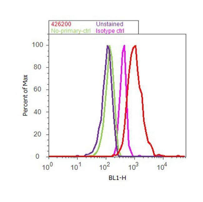
Supportive validation
- Submitted by
- Invitrogen Antibodies (provider)
- Main image

- Experimental details
- Flow cytometry analysis of CAP43 was done on HEK-293 cells. Cells were fixed with 70% ethanol for 10 minutes, permeabilized with 0.25% Triton™ X-100 for 20 minutes, and blocked with 5% BSA for 30 minutes at room temperature. Cells were labeled with CAP43 Rabbit Polyclonal Antibody (42-6200, red histogram) or with rabbit isotype control (pink histogram) at 3-5 ug/million cells in 2.5% BSA. After incubation at room temperature for 2 hours, the cells were labeled with Alexa Fluor® 488 Goat Anti-Rabbit Secondary Antibody (A11008) at a dilution of 1:400 for 30 minutes at room temperature. The representative 10, 000 cells were acquired and analyzed for each sample using an Attune® Acoustic Focusing Cytometer. The purple histogram represents unstained control cells and the green histogram represents no-primary-antibody control.