Explore
 Validate
 Learn
Learn Western blot
Antibody data
- Antibody Data
- Antigen structure
- References [2]
- Comments [0]
- Validations
- Western blot [3]
- Immunohistochemistry [3]
- Other assay [3]
Submit
Validation data
Reference
Comment
Report error
- Product number
- PA5-18109 - Provider product page

- Provider
- Invitrogen Antibodies
- Product name
- NDRG1 Polyclonal Antibody
- Antibody type
- Polyclonal
- Antigen
- Synthetic peptide
- Description
- This antibody is predicted to react with canine, mouse and rat based on sequence homology. This antibody is tested in Peptide ELISA: antibody detection limit dilution 32,000.
- Reactivity
- Human
- Host
- Goat
- Isotype
- IgG
- Vial size
- 100 µg
- Concentration
- 0.5 mg/mL
- Storage
- -20° C, Avoid Freeze/Thaw Cycles
Submitted references Cell and context-dependent sorting of neuropathy-associated protein NDRG1 - insights from canine tissues and primary Schwann cell cultures.
N-myc downstream-regulated gene 1 inhibits the proliferation and invasion of hepatocellular carcinoma cells via the regulation of integrin β3.
Skedsmo FS, Tranulis MA, Espenes A, Prydz K, Matiasek K, Gunnes G, Hermansen LC, Jäderlund KH
BMC veterinary research 2019 Apr 27;15(1):121
BMC veterinary research 2019 Apr 27;15(1):121
N-myc downstream-regulated gene 1 inhibits the proliferation and invasion of hepatocellular carcinoma cells via the regulation of integrin β3.
Song Y, Wu G, Zhang M, Kong Q, Du J, Zheng Y, Yue L, Cao L
Oncology letters 2017 May;13(5):3599-3607
Oncology letters 2017 May;13(5):3599-3607
No comments: Submit comment
Supportive validation
- Submitted by
- Invitrogen Antibodies (provider)
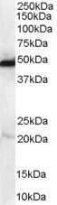
- Main image

- Experimental details
- Western Blot staining of Human Kidney lysate using Product # PA5-18109 at a concentration of 0.1 µg/mL, the primary antibody incubation was 1 hour and the detection method was chemiluminescence.
- Submitted by
- Invitrogen Antibodies (provider)
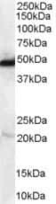
- Main image

- Experimental details
- Western Blot staining of Human Kidney lysate using Product # PA5-18109 at a concentration of 0.1 µg/mL, the primary antibody incubation was 1 hour and the detection method was chemiluminescence.
- Submitted by
- Invitrogen Antibodies (provider)
- Main image
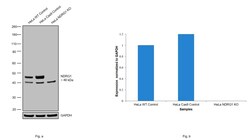
- Experimental details
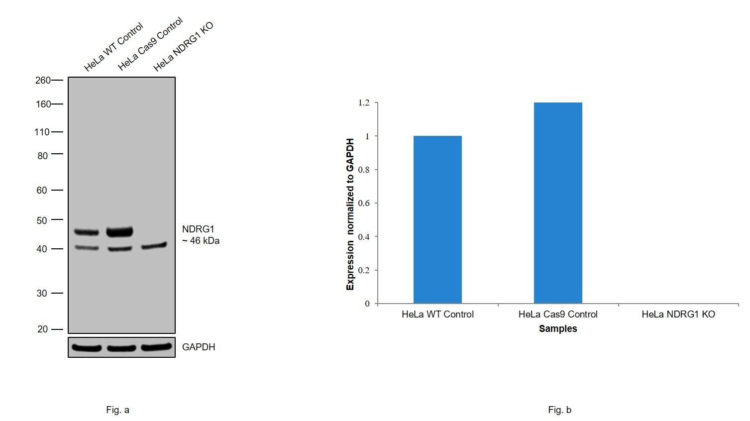
- Knockout of NDRG1 was achieved by CRISPR-Cas9 genome editing using LentiArray™ Lentiviral sgRNA (Product # A32042, Assay ID CRISPR1035265_LV) and LentiArray Cas9 Lentivirus (Product # A32064). Western blot analysis of NDRG1 was performed by loading 30 µg of HeLa Wild Type (Lane 1), HeLa CAS9 (Lane 2) and HeLa NDRG1 KO (Lane 3) whole cell extracts. The samples were electrophoresed using NuPAGE™ Novex™ 4-12% Bis-Tris Protein Gel (Product # NP0322BOX). Resolved proteins were then transferred onto a nitrocellulose membrane (Product # IB23001) by iBlot® 2 Dry Blotting System (Product # IB21001). The blot was probed with Anti NDRG1 Polyclonal Antibody (Product # PA5-18109, 0.2 µg/mL dilution) and Rabbit anti-Goat IgG (H+L) Superclonal™ Recombinant Secondary Antibody, HRP (Product # A27014, 1:4000 dilution) using the iBright FL 1000 (Product # A32752). Chemiluminescent detection was performed using Novex® ECL Chemiluminescent Substrate Reagent Kit (Product # WP20005). Loss of signal upon CRISPR mediated knockout (KO) using the LentiArray™ CRISPR product line confirms that antibody is specific to NDRG1. Uncharacterized band at ~ 41 kDa was observed in all the samples.
Supportive validation
- Submitted by
- Invitrogen Antibodies (provider)
- Main image

- Experimental details
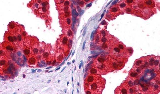
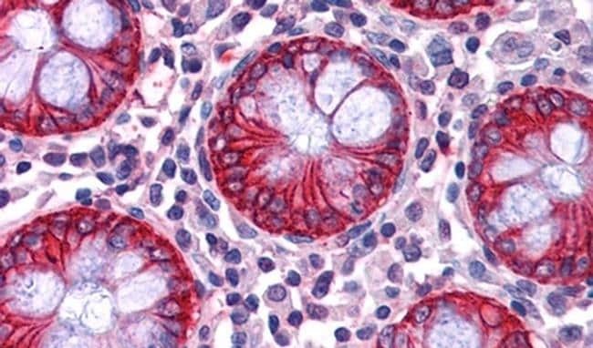
- Immunohistochemical analysis of NDRG1 in Human Prostate using a NDRG1 monoclonal antibody (Product #PA5-18109) at 2.5 µg/mL. The Human Prostate tissue section was paraffin embeded and detected using steamed antigen retrieval with citrate buffer pH 6, AP-staining.
- Submitted by
- Invitrogen Antibodies (provider)
- Main image
- Experimental details
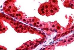
- Immunohistochemical staining of paraffin embedded of Human Prostate using Product # PA5-18109 at a concentration of 2.5 µg/mL. The tissue was processed by steamed antigen retrieval with citrate buffer pH 6 and stained with AP.
- Submitted by
- Invitrogen Antibodies (provider)
- Main image
- Experimental details
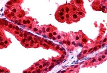
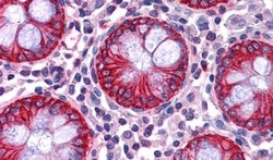
- Immunohistochemical analysis of NDRG1 in Human Colon using a NDRG1 monoclonal antibody (Product #PA5-18109) at 2.5 µg/mL. The Human Colon tissue section was paraffin embeded and detected using steamed antigen retrieval with citrate buffer pH 6, AP-staining.
Supportive validation
- Submitted by
- Invitrogen Antibodies (provider)
- Main image
- Experimental details
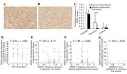
- NULL

- Submitted by
- Invitrogen Antibodies (provider)
- Main image
- Experimental details
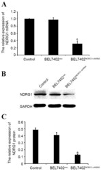
- Figure 3. NDRG1-shRNA interfering efficiency in BEL7402 cells. (A) Quantitative polymerase chain reaction detected NDRG1-shRNA interfering efficiency in BEL7402 cells. (B) Western blot analysis of NDRG1 protein expression levels in BEL7402 vec , BEL7402 NDRG1-shRNA and control cells. (C) NDRG1 protein expression histogram in BEL7402 vec , BEL7402 NDRG1-shRNA and control cells. *P

- Submitted by
- Invitrogen Antibodies (provider)
- Main image

- Experimental details
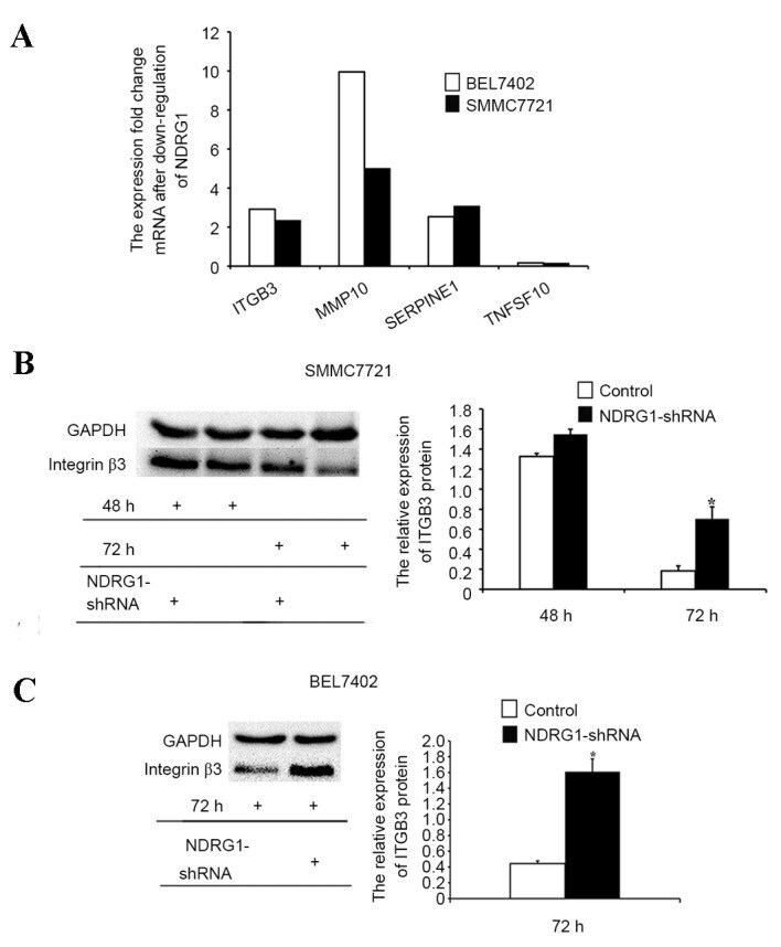
- Figure 6. Results of polymerase chain reaction demonstrating that the downregulation of NDRG1 in BEL7402 and SMMC7721 cell lines resulted in subsequent up- or downregulation of different genes. (A) Effect of NDRG1 on the expression levels of ITGB3, MMP10, SERPINE1 and TNFSF10 in the BEL7402 and SMMC7721 cell lines. ITGB3 protein expression was detected by western blot analysis prior to and subsequent to the downregulation of the NDRG1 gene in (B) BEL7402 and (C) SMMC7721 cells. *P