Explore
 Validate
 Learn
Learn Western blot
Antibody data
- Antibody Data
- Antigen structure
- References [0]
- Comments [0]
- Validations
- Western blot [4]
- Immunohistochemistry [1]
Submit
Validation data
Reference
Comment
Report error
- Product number
- PA5-27213 - Provider product page

- Provider
- Invitrogen Antibodies
- Product name
- NDRG1 Polyclonal Antibody
- Antibody type
- Polyclonal
- Antigen
- Recombinant protein fragment
- Description
- Recommended positive controls: 293T, HepG2, NIH-3T3. Predicted reactivity: Mouse (98%), Rat (98%), Xenopus laevis (85%), Dog (98%), Bovine (98%). Store product as a concentrated solution. Centrifuge briefly prior to opening the vial.
- Reactivity
- Human, Mouse
- Host
- Rabbit
- Isotype
- IgG
- Vial size
- 100 µL
- Concentration
- 0.67 mg/mL
- Storage
- Store at 4°C short term. For long term storage, store at -20°C, avoiding freeze/thaw cycles.
No comments: Submit comment
Supportive validation
- Submitted by
- Invitrogen Antibodies (provider)
- Main image

- Experimental details
- Western Blot using NDRG1 Polyclonal Antibody (Product # PA5-27213). Sample (30 µg of whole cell lysate). A: NIH-3T3. 10% SDS PAGE. NDRG1 Polyclonal Antibody (Product # PA5-27213) diluted at 1:1,000.

- Submitted by
- Invitrogen Antibodies (provider)
- Main image

- Experimental details
- NDRG1 Polyclonal Antibody detects NDRG1 protein by Western blot analysis. A. 30 µg 293T whole cell lysate/extract. B. 30 µg HepG2 whole cell lysate/extract.10 % SDS-PAGE. NDRG1 Polyclonal Antibody (Product # PA5-27213) dilution: 1:1,000.

- Submitted by
- Invitrogen Antibodies (provider)
- Main image

- Experimental details
- Western Blot using NDRG1 Polyclonal Antibody (Product # PA5-27213). Whole cell extract (30 µg) was separated by 10% SDS-PAGE, and the membrane was blotted with NDRG1 Polyclonal Antibody (Product # PA5-27213) diluted at 1:1,000. The HRP-conjugated anti-rabbit IgG antibody was used to detect the primary antibody, and the signal was developed with Trident ECL plus-Enhanced.

- Submitted by
- Invitrogen Antibodies (provider)
- Main image

- Experimental details
- Knockout of NDRG1 was achieved by CRISPR-Cas9 genome editing using LentiArray™ Lentiviral sgRNA (Product # A32042, Assay ID CRISPR1035265_LV) and LentiArray Cas9 Lentivirus (Product # A32064). Western blot analysis of NDRG1 was performed by loading 30 µg of HeLa wild type (Lane 1), HeLa Cas9 (Lane 2) and HeLa NDRG1 KO (Lane 3) whole cell extracts. The samples were electrophoresed using NuPAGE™ Novex™ 4-12% Bis-Tris Protein Gel (Product # NP0322BOX). Resolved proteins were then transferred onto a nitrocellulose membrane (Product # IB23001) by iBlot® 2 Dry Blotting System (Product # IB21001). The blot was probed with Anti-NDRG1 Polyclonal Antibody (Product # PA5-27213, 1:2000 dilution) and Goat anti-Rabbit IgG (H+L) Superclonal™ Recombinant Secondary Antibody, HRP (Product # A27036, 1:4000 dilution) using the iBright FL 1000 (Product # A32752). Chemiluminescent detection was performed using Novex® ECL Chemiluminescent Substrate Reagent Kit (Product # WP20005). Loss of signal upon CRISPR mediated knockout (KO) using the LentiArray™ CRISPR product line confirms that antibody is specific to NDRG1.

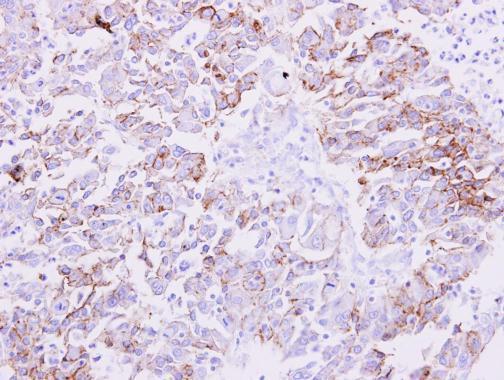
Supportive validation
- Submitted by
- Invitrogen Antibodies (provider)
- Main image

- Experimental details
- NDRG1 Polyclonal Antibody detects NDRG1 protein at membrane on human liver carcinoma by immunohistochemical analysis. Sample: Paraffin-embedded liver carcinoma. NDRG1 Polyclonal Antibody (Product # PA5-27213) dilution: 1:500. Antigen Retrieval: EDTA based buffer, pH 8.0, 15 min.