Explore
 Validate
 Learn
Learn Western blot
 Immunocytochemistry
Antibody data
- Antibody Data
- Antigen structure
- References [13]
- Comments [0]
- Validations
- Immunocytochemistry [1]
- Immunohistochemistry [1]
Submit
Validation data
Reference
Comment
Report error
- Product number
- HPA006881 - Provider product page

- Provider
- Atlas Antibodies
- Proper citation
- Atlas Antibodies Cat#HPA006881, RRID:AB_1078392
- Product name
- Anti-NDRG1
- Antibody type
- Polyclonal
- Description
- Polyclonal Antibody against Human NDRG1, Gene description: N-myc downstream regulated 1, Alternative Gene Names: CAP43, DRG1, NDR1, RTP, TDD5, Validated applications: WB, IHC, ICC, Uniprot ID: Q92597, Storage: Store at +4°C for short term storage. Long time storage is recommended at -20°C.
- Reactivity
- Human, Rat
- Host
- Rabbit
- Conjugate
- Unconjugated
- Isotype
- IgG
- Vial size
- 100 µl
- Concentration
- 0.1 mg/ml
- Storage
- Store at +4°C for short term storage. Long time storage is recommended at -20°C.
- Handling
- The antibody solution should be gently mixed before use.
Submitted references NDRG1 facilitates self-renewal of liver cancer stem cells by preventing EpCAM ubiquitination
N-myc downstream regulated gene 1 (ndrg1) functions as a molecular switch for cellular adaptation to hypoxia
Identification of PIM1 substrates reveals a role for NDRG1 phosphorylation in prostate cancer cellular migration and invasion
Differential expression and hypoxia‐mediated regulation of the N‐myc downstream regulated gene family
Identification and Characterization of Cancer Cells That Initiate Metastases to the Brain and Other Organs
The up‐regulation of NDRG1 by HIF counteracts the cancer‐promoting effect of HIF in VHL‐deficient clear cell renal cell carcinoma
Cell and context-dependent sorting of neuropathy-associated protein NDRG1 – insights from canine tissues and primary Schwann cell cultures
Hypoxia Inducible Factor 1α Inhibits the Expression of Immunosuppressive Tryptophan-2,3-Dioxygenase in Glioblastoma
The metastatic suppressor NDRG1 inhibits EMT, migration and invasion through interaction and promotion of caveolin-1 ubiquitylation in human colorectal cancer cells
The Metastasis Suppressor, N-myc Downregulated Gene 1 (NDRG1), Is a Prognostic Biomarker for Human Colorectal Cancer
N-myc Downstream Regulated 1 (NDRG1) Is Regulated by Eukaryotic Initiation Factor 3a (eIF3a) during Cellular Stress Caused by Iron Depletion
A Deletion in the N-Myc Downstream Regulated Gene 1 (NDRG1) Gene in Greyhounds with Polyneuropathy
NDRG4 Is Required for Cell Cycle Progression and Survival in Glioblastoma Cells
Cheng Q, Ning S, Zhu L, Zhang C, Jiang S, Hao Y, Zhu J
British Journal of Cancer 2023;129(2):237-248
British Journal of Cancer 2023;129(2):237-248
N-myc downstream regulated gene 1 (ndrg1) functions as a molecular switch for cellular adaptation to hypoxia
Park J, Gabel A, Kassir P, Kang L, Chowdhary P, Osei-Ntansah A, Tran N, Viswanathan S, Canales B, Ding P, Lee Y, Brewster R
eLife 2022;11
eLife 2022;11
Identification of PIM1 substrates reveals a role for NDRG1 phosphorylation in prostate cancer cellular migration and invasion
Ledet R, Ruff S, Wang Y, Nayak S, Schneider J, Ueberheide B, Logan S, Garabedian M
Communications Biology 2021;4(1)
Communications Biology 2021;4(1)
Differential expression and hypoxia‐mediated regulation of the N‐myc downstream regulated gene family
Le N, Hufford T, Park J, Brewster R
The FASEB Journal 2021;35(11)
The FASEB Journal 2021;35(11)
Identification and Characterization of Cancer Cells That Initiate Metastases to the Brain and Other Organs
Berghoff A, Liao Y, Karreman M, Ilhan-Mutlu A, Gunkel K, Sprick M, Eisen C, Kessler T, Osswald M, Wünsche S, Feinauer M, Gril B, Marmé F, Michel L, Bago-Horvath Z, Sahm F, Becker N, Breckwoldt M, Solecki G, Gömmel M, Huang L, Rübmann P, Thome C, Ratliff M, Trumpp A, Steeg P, Preusser M, Wick W, Winkler F
Molecular Cancer Research 2021;19(4):688-701
Molecular Cancer Research 2021;19(4):688-701
The up‐regulation of NDRG1 by HIF counteracts the cancer‐promoting effect of HIF in VHL‐deficient clear cell renal cell carcinoma
Zhang Z, Zhang S, Chen H, Mao Y, Li Z, Kong C, Han B, Zhang J, Chen Y, Xue W, Zhai W, Wang L
Cell Proliferation 2020;53(7)
Cell Proliferation 2020;53(7)
Cell and context-dependent sorting of neuropathy-associated protein NDRG1 – insights from canine tissues and primary Schwann cell cultures
Skedsmo F, Tranulis M, Espenes A, Prydz K, Matiasek K, Gunnes G, Hermansen L, Jäderlund K
BMC Veterinary Research 2019;15(1)
BMC Veterinary Research 2019;15(1)
Hypoxia Inducible Factor 1α Inhibits the Expression of Immunosuppressive Tryptophan-2,3-Dioxygenase in Glioblastoma
Mohapatra S, Sadik A, Tykocinski L, Dietze J, Poschet G, Heiland I, Opitz C
Frontiers in Immunology 2019;10
Frontiers in Immunology 2019;10
The metastatic suppressor NDRG1 inhibits EMT, migration and invasion through interaction and promotion of caveolin-1 ubiquitylation in human colorectal cancer cells
Mi L, Zhu F, Yang X, Lu J, Zheng Y, Zhao Q, Wen X, Lu A, Wang M, Zheng M, Ji J, Sun J
Oncogene 2017;36(30):4323-4335
Oncogene 2017;36(30):4323-4335
The Metastasis Suppressor, N-myc Downregulated Gene 1 (NDRG1), Is a Prognostic Biomarker for Human Colorectal Cancer
Lo A, Mao Z, Sun J, Feng B, Ma J, Zang L, Dong F, Zhang D, Zheng M
PLoS ONE 2013;8(7):e68206
PLoS ONE 2013;8(7):e68206
N-myc Downstream Regulated 1 (NDRG1) Is Regulated by Eukaryotic Initiation Factor 3a (eIF3a) during Cellular Stress Caused by Iron Depletion
Zhang J, Lane D, Saletta F, Rahmanto Y, Kovacevic Z, Richardson D
PLoS ONE 2013;8(2):e57273
PLoS ONE 2013;8(2):e57273
A Deletion in the N-Myc Downstream Regulated Gene 1 (NDRG1) Gene in Greyhounds with Polyneuropathy
Lewin A, Drögemüller C, Becker D, Kessler B, Kemter E, Tetens J, Jurina K, Hultin Jäderlund K, Flagstad A, Perloski M, Lindblad-Toh K, Matiasek K
PLoS ONE 2010;5(6):e11258
PLoS ONE 2010;5(6):e11258
NDRG4 Is Required for Cell Cycle Progression and Survival in Glioblastoma Cells
Schilling S, Hjelmeland A, Radiloff D, Liu I, Wakeman T, Fielhauer J, Foster E, Lathia J, Rich J, Wang X, Datto M
Journal of Biological Chemistry 2009;284(37):25160-25169
Journal of Biological Chemistry 2009;284(37):25160-25169
No comments: Submit comment
Supportive validation
- Submitted by
- Atlas Antibodies (provider)
- Main image

- Experimental details
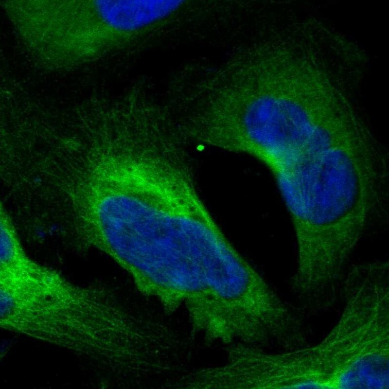
- Immunofluorescent staining of human cell line U-2 OS shows localization to cytosol & microtubules.
- Sample type
- Human
Supportive validation
- Submitted by
- Atlas Antibodies (provider)
- Enhanced method
- Orthogonal validation
- Main image
- Experimental details
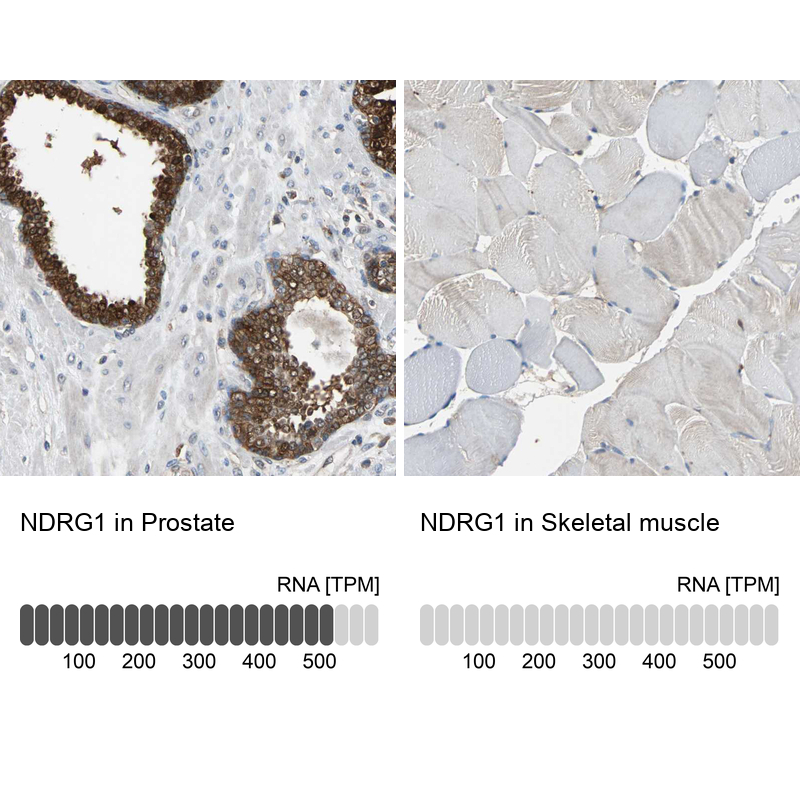
- Immunohistochemistry analysis in human prostate and skeletal muscle tissues using HPA006881 antibody. Corresponding NDRG1 RNA-seq data are presented for the same tissues.
- Sample type
- Human
- Protocol
- Protocol