Explore
 Validate
 Learn
Learn Western blot
Antibody data
- Antibody Data
- Antigen structure
- References [3]
- Comments [0]
- Validations
- Western blot [1]
- Immunohistochemistry [2]
- Other assay [3]
Submit
Validation data
Reference
Comment
Report error
- Product number
- PA5-34517 - Provider product page

- Provider
- Invitrogen Antibodies
- Product name
- AGR2 Polyclonal Antibody
- Antibody type
- Polyclonal
- Antigen
- Synthetic peptide
- Description
- A suggested positive control is Hela cell lysate. PA5-34517 can be used with blocking peptide PEP-1560.
- Reactivity
- Human
- Host
- Rabbit
- Isotype
- IgG
- Vial size
- 100 µg
- Concentration
- 1 mg/mL
- Storage
- Maintain refrigerated at 2-8°C for up to 3 months. For long term storage store at -20°C
Submitted references Serum Level of Tumor-Overexpressed AGR2 Is Significantly Associated with Unfavorable Prognosis of Canine Malignant Mammary Tumors.
Building a Thick Mucus Hydrogel Layer to Improve the Physiological Relevance of In Vitro Primary Colonic Epithelial Models.
Transitional basal cells at the squamous-columnar junction generate Barrett's oesophagus.
Yuan SH, Chang SC, Huang Y, Liu HP
Animals : an open access journal from MDPI 2021 Oct 9;11(10)
Animals : an open access journal from MDPI 2021 Oct 9;11(10)
Building a Thick Mucus Hydrogel Layer to Improve the Physiological Relevance of In Vitro Primary Colonic Epithelial Models.
Wang Y, Kim R, Sims CE, Allbritton NL
Cellular and molecular gastroenterology and hepatology 2019;8(4):653-655.e5
Cellular and molecular gastroenterology and hepatology 2019;8(4):653-655.e5
Transitional basal cells at the squamous-columnar junction generate Barrett's oesophagus.
Jiang M, Li H, Zhang Y, Yang Y, Lu R, Liu K, Lin S, Lan X, Wang H, Wu H, Zhu J, Zhou Z, Xu J, Lee DK, Zhang L, Lee YC, Yuan J, Abrams JA, Wang TC, Sepulveda AR, Wu Q, Chen H, Sun X, She J, Chen X, Que J
Nature 2017 Oct 26;550(7677):529-533
Nature 2017 Oct 26;550(7677):529-533
No comments: Submit comment
Supportive validation
- Submitted by
- Invitrogen Antibodies (provider)
- Main image

- Experimental details
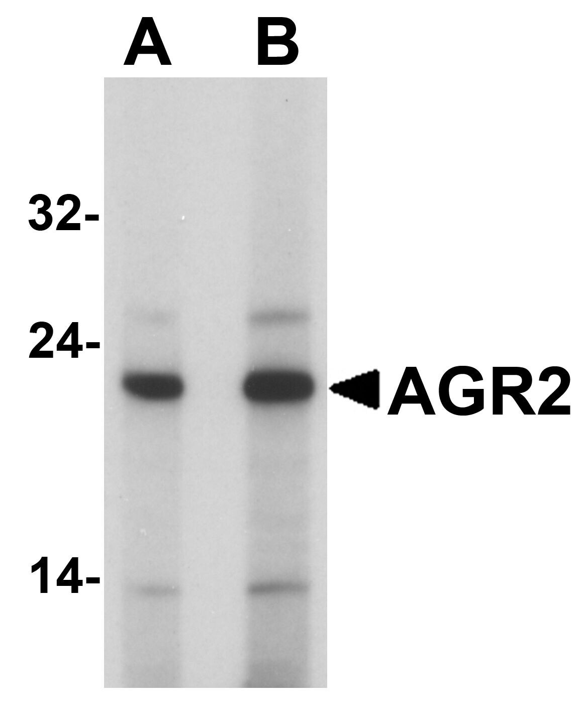
- Western Blot analysis of AGR2 in Hela cell lysate with AGR2 Polyclonal Antibody (Product # PA5-34517) at (A) 1 and (B) 2 µg/mL
Supportive validation
- Submitted by
- Invitrogen Antibodies (provider)
- Main image
- Experimental details
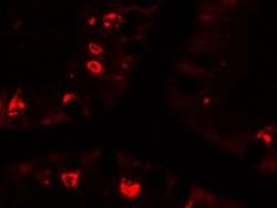
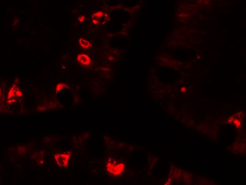
- Immunofluorescence of AGR2 in human small intestine tissue with AGR2 Polyclonal Antibody (Product # PA5-34517) at 20 µg/mL.
- Submitted by
- Invitrogen Antibodies (provider)
- Main image
- Experimental details
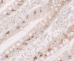
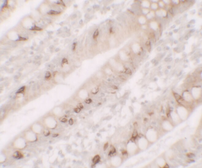
- Immunohistochemistry of AGR2 in human small intestine tissue with AGR2 Polyclonal Antibody (Product # PA5-34517) at 5 µg/mL.
Supportive validation
- Submitted by
- Invitrogen Antibodies (provider)
- Main image
- Experimental details
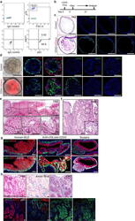
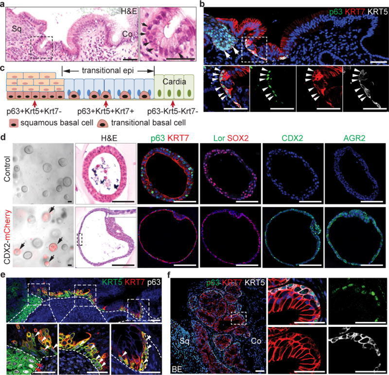
- Extended Data Figure 11 Different response to CDX2 overexpression in the transitional (p63 + KRT7 + ) and the squamous (p63 + KRT7 - ) basal progenitor cells in vitro. Human and mouse MLE present similar gene expression a , Two distinct basal progenitor populations (p63 + KRT7 - Vs p63 + KRT7 + ) are present at the human SCJ as indicated by flow cytometric analysis. n =3 independent experiments. b , Schematic depicts the induction of CDX2 overexpression with Doxycycline treatment of CDX2 virus-infected human SCJ basal progenitor cells. c , CDX2 overexpression promotes intestinal metaplasia of p63 + KRT7 + cells. The metaplastic columnar cells are PAS + and express Villin1, Muc2 and TFF3. n =6 per group. d , Ectopic CDX2 expression does not promote intestinal metaplasia of the stratified squamous epithelium in organoids formed by p63 + KRT7 - squamous basal cells. n =4 per group. e , The transitional epithelium with underlying basal cells is dramatically expanded in patients with long-term gastro-esophageal acid reflux. Dotted lines indicate the basement membrane. n =3. f , The transitional epithelium with basal cells is amplified in BE mixed with MLE. n =5. g , Similar phenotypic presentation of human MLE and mouse MLE developed at the SCJ following CDX2 overexpression and oesophageal-duodenal anastomosis surgery. human MLE n =10; Krt5-rtTA; otet-CDX2 mutants n =5; surgical mice n =5. h , Goblet cells in human BE is positive for Alcian blue and

- Submitted by
- Invitrogen Antibodies (provider)
- Main image
- Experimental details
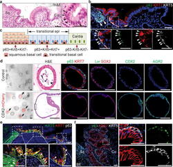
- Figure 4 The transitional epithelium is also present in the human SCJ and amplified during BE pathogenesis a , The transitional epithelium consists of basal progenitors (arrows) and luminal cells. n =5. b , Basal progenitor cells in the transitional epithelium express p63, KRT5 and KRT7 (arrowheads). Note that basal cells of the stratified squamous (sq) cells are KRT7 - (arrows). n =5. c , Schematic shows two types of basal progenitor cells in the human SCJ. d , CDX2 overexpression promotes intestinal metaplasia of p63 + KRT7 + cells in 3D organoid culture. n =6 per group. e , The transitional epithelium with underlying basal cells is dramatically expanded in patients with gastro-oesophageal acid reflux. Dotted lines indicate the basement membrane. n =3. f , The transitional epithelium with basal cells is amplified in BE mixed with MLE. n =5. Me, mesenchyme. Scale bar, 50 mum.
- Submitted by
- Invitrogen Antibodies (provider)
- Main image
- Experimental details
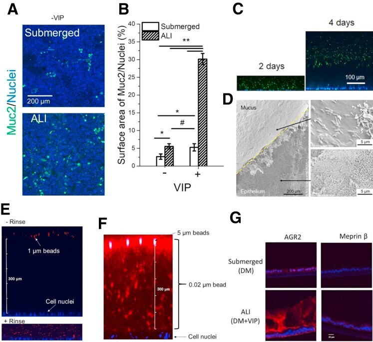
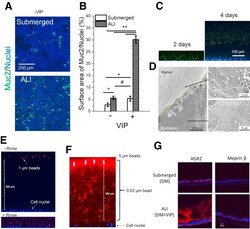
- Supplemental Figure 2 ( A , B ) Effect of culture method and VIP (0 and 330 ng/mL) on the number of goblet cells. ( A ) Immunofluorescence images of monolayers stained for Muc2 (green). ( B ) The percentage of the monolayer surface area positive for Muc 2 fluorescence. * P < .05; ** P < .005; #not statistically significant. n = 3. ( C , D ) The hydrated mucus layer generated by VIP-assisted air-liquid interface (ALI) culture. ( C ) Side-view confocal micrograph showing tissues with bacteria-separating mucus accumulation at 2 and 4 days, respectively. Green: GFP-expressing EC; blue: Hoechst 33342. ( D ) Scanning electron microscopy of hydrated mucus layer (overlaid with GFP-expressing EC). The mucus layer was partially removed using tweezers to reveal the epithelium (lower arrow) below the mucus. The upper right panel shows bacteria (rod-shaped structures) on the surface of the mucus layer. ( E-G ) Characterizations of the hydrated mucus layer generated by VIP-assisted ALI culture. ( E ) Visualization of the mucus layer by overlaying 1-mum red fluorescent beads. Top: adding beads to the mucus layer without rinsing. Bottom: adding beads to the mucus layer after gently rinsing. ( F ) The mucus layer was overlaid with a mixture of 0.02-mum red fluorescent beads (#F8786; ThermoFisher) and 5_mum green fluorescent beads (#G0500; ThermoFisher) for 4 hours. ( G ) Immunofluorescence of sectioned monolayers. Red: AGR2 or Meprin beta; blue: Hoechst 33342. There was minimal Meprin beta ex