 Explore
 Validate
 Learn
Learn Western blot
Antibody data
- Antibody Data
- Antigen structure
- References [1]
- Comments [0]
- Validations
- Western blot [2]
- Immunohistochemistry [5]
- Other assay [4]
Submit
Validation data
Reference
Comment
Report error
- Product number
- MA5-16244 - Provider product page

- Provider
- Invitrogen Antibodies
- Product name
- AGR2 Monoclonal Antibody (10E2)
- Antibody type
- Monoclonal
- Antigen
- Other
- Reactivity
- Human, Mouse, Rat, Bovine, Canine
- Host
- Mouse
- Isotype
- IgG
- Antibody clone number
- 10E2
- Vial size
- 100 µg
- Concentration
- 0.5 mg/mL
- Storage
- Store at 4°C short term. For long term storage, store at -20°C, avoiding freeze/thaw cycles.
Submitted references Serum Level of Tumor-Overexpressed AGR2 Is Significantly Associated with Unfavorable Prognosis of Canine Malignant Mammary Tumors.
Yuan SH, Chang SC, Huang Y, Liu HP
Animals : an open access journal from MDPI 2021 Oct 9;11(10)
Animals : an open access journal from MDPI 2021 Oct 9;11(10)
No comments: Submit comment
Supportive validation
- Submitted by
- Invitrogen Antibodies (provider)
- Main image

- Experimental details
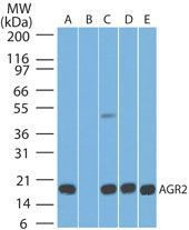
- Western blot analysis of AGR2 in A) human stomach tissue lysate in the absense of immunizing peptide, B) human stomach tissue lysate in the presence of immunizing peptide, C) mouse stomach tissue lysate, D) rat stomach tissue lysate, and E) HCT-116 cell lysate using a AGR 2 monoclonal antibody (Product # MA5-16244) at 5 µg/mL. Goat anti-mouse Ig HRP secondary antibody.
- Submitted by
- Invitrogen Antibodies (provider)
- Main image
- Experimental details
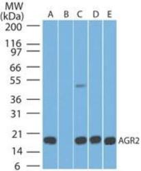
- Western blot analysis of AGR2 in human stomach. Samples were incubated in AGR2 monoclonal antibody (Product # MA5-16244) using a dilution of 5 µg/mL followed by a goat anti-mouse Ig HRP secondary antibody. A) absense and B) presence of immunizing peptide, C) mouse stomach, D) rat stomach tissue lysate, and E) HCT-116 cell lysate. PicoTect ECL substrate solution was used for this test.

Supportive validation
- Submitted by
- Invitrogen Antibodies (provider)
- Main image
- Experimental details
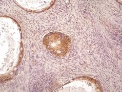
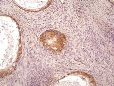
- Immunohistochemical analysis of AGR2 in a human ovarian cancer section. Samples were incubated in AGR2 monoclonal antibody (Product # MA5-16244) using a dilution of 5 µg/mL. The representative image shows AGR2 immuno-reactivity in the developing ovarian cancer and the follicle cells of Graafian follicles. The cells of medulla region in the ovary did not show AGR2 positivity. [10X magnification].
- Submitted by
- Invitrogen Antibodies (provider)
- Main image

- Experimental details
- Immunohistochemical analysis of AGR2 in a section of human bladded cancer. Samples were incubated in AGR2 monoclonal antibody (Product # MA5-16244) using a dilution of 5 µg/mL. The AGR2 immunoreactivity was observed in cellular cytoplasm and the inter-cellular spaces in cancerous areas of the sections. [40X magnification].

- Submitted by
- Invitrogen Antibodies (provider)
- Main image

- Experimental details
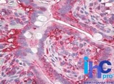
- Immunohistochemical analysis of AGR2 in paraffin-embedded human small intestine tissue. Samples were incubated in AGR2 monoclonal antibody (Product # MA5-16244) using a dilution of 10 µg/mL. Staining of formalin-fixed tissues is enhanced by boiling tissue sections in 10 mM sodium citrate buffer, pH 6.0 for 10-20 min followed by cooling at RT for 20 min.
- Submitted by
- Invitrogen Antibodies (provider)
- Main image

- Experimental details
- Immunohistochemical analysis of AGR2 in formalin-fixed, paraffin-embedded human breast. Samples were incubated in AGR2 monoclonal antibody (Product # MA5-16244) using a dilution of 5 µg/mL. Staining of formalin-fixed tissues is enhanced by boiling tissue sections in 10 mM sodium citrate buffer, pH 6.0 for 10-20 min followed by cooling at RT for 20 min.

- Submitted by
- Invitrogen Antibodies (provider)
- Main image

- Experimental details
- Immunohistochemical analysis of AGR2 in formalin-fixed, paraffin-embedded human spleen tissue. Samples were incubated in AGR2 monoclonal antibody (Product # MA5-16244) using a dilution of 5 µg/mL.

Supportive validation
- Submitted by
- Invitrogen Antibodies (provider)
- Main image

- Experimental details
- Figure 1 AGR2 overexpression in canine MMT tissues revealed by immunohistochemistry (IHC). AGR2 protein expression was analyzed with tissue slides of 34 canine MMT cases using an antibody specific to AGR2. ( A ) Micrographs of two representative cases. Upper micrographs were acquired at 100x magnification; bottom micrographs were the areas in the upper images acquired at 400x magnification. ( B ) MMT tissues stained with an isotype control IgG. ( C ) Pairwise comparison of IHC scores of AGR2 between MMT tissue and adjacent normal mammary gland tissue of the same patient. Each line represents an individual patient. Statistical significance was determined by two-tailed paired t -test. ( D ) Comparison of IHC scores of AGR2 between overall MMT tissues and adjacent normal mammary tissues. L, M, and H denote low, moderate, and high levels of AGR2, respectively. Statistical significance was determined by Fisher's exact test. ** p < 0.01; *** p < 0.001.

- Submitted by
- Invitrogen Antibodies (provider)
- Main image
- Experimental details
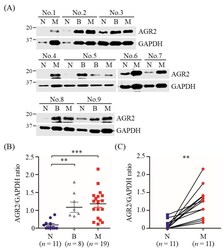
- Figure 2 Elevated protein levels of AGR2 in canine MMT tissues compared with paired normal mammary tissues. Tissue homogenates (20 ug proteins each sample) of MMT (denoted M), paired with benign mammary tumor (denoted B) and normal mammary gland tissues (denoted N) collected from the same patient, were analyzed by immunoblotting using an antibody specific to AGR2. GAPDH was used as an internal control. Representative results of nine cases (No. 1 to No. 9) were shown in ( A ). AGR2 protein levels in individual samples were determined by calculating the AGR2-to-GAPDH ratio. ( B ) Comparison of AGR2 protein levels between tissue types. Statistical significance was determined by Mann-Whitney U test. ( C ) Pairwise comparison of AGR2 protein levels between MMT and paired normal mammary gland tissues of the same patient. Lines represent individual patients. Statistical significance was determined by two-tailed paired t -test. ** p < 0.01; *** p < 0.001.

- Submitted by
- Invitrogen Antibodies (provider)
- Main image

- Experimental details
- Figure 3 Detection of AGR2 expression in canine MMT cells and conditioned media. ( A ) Intracellular localization of AGR2 in canine MMT cell lines, DMGT, and CF41.Mg. AGR2 expression was verified by immunofluorescence staining using an AGR2-specific antibody. DAPI staining indicated the nuclei. Magnification, x40. ( B ) Detection of AGR2 in cell lysates of canine MMT cell lines. Cell lysates (30 ug proteins per sample) were analyzed by immunoblotting with an AGR2-specific antibody. ( C ) Detection of extracellular AGR2 (eAGR2) in serum-free conditioned media of canine MMT cells. Cells were grown in serum-free media for one day, and the conditioned media were harvested and subsequently concentrated. The resulting conditioned media (50 ug proteins per sample) were analyzed using a competitive ELISA established for eAGR2 detection.

- Submitted by
- Invitrogen Antibodies (provider)
- Main image
- Experimental details
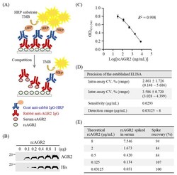
- Figure 4 Establishment of a competitive ELISA for detection of serum eAGR2 in MMT dogs. ( A ) Scheme for the established competitive ELISA. ( B ) Identity of recombinant canine AGR2 (rcAGR2) was confirmed by immunoblotting using antibodies specific to AGR2 and 6xHis tag, respectively. ( C ) A standard curve with fourfold serially diluted rcAGR2 proteins (8000, 2000, 500, 125, and 31.25 ng/mL) was set for the competitive ELISA. Data are presented as the mean values +- SD across 10 assays. ( D ) Precision of the competitive ELISA. The intra-assay coefficient variant (CV), inter-assay CV, and the detection sensitivity were determined across 10 assays. ( E ) Evaluation of serum influence on eAGR2 detection in the ELISA. A spike recovery test was performed by detecting rcAGR2 spiked at indicated concentrations into canine serum containing undetectable eAGR2. The recovery rate (%) of individual spike rcAGR2 was calculated as indicated.
