Explore
 Validate
 Learn
Learn Western blot
Antibody data
- Antibody Data
- Antigen structure
- References [1]
- Comments [0]
- Validations
- Western blot [2]
- Immunocytochemistry [2]
- Immunohistochemistry [2]
Submit
Validation data
Reference
Comment
Report error
- Product number
- GTX113629 - Provider product page

- Provider
- GeneTex
- Proper citation
- GeneTex Cat#GTX113629, RRID:AB_2036281
- Product name
- ASL antibody
- Antibody type
- Polyclonal
- Reactivity
- Human, Mouse
- Host
- Rabbit
Submitted references Understanding the role of argininosuccinate lyase transcript variants in the clinical and biochemical variability of the urea cycle disorder argininosuccinic aciduria.
Hu L, Pandey AV, Eggimann S, Rüfenacht V, Möslinger D, Nuoffer JM, Häberle J
The Journal of biological chemistry 2013 Nov 29;288(48):34599-611
The Journal of biological chemistry 2013 Nov 29;288(48):34599-611
No comments: Submit comment
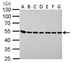
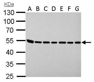
Supportive validation
- Submitted by
- GeneTex (provider)
- Main image
- Experimental details
- ASL antibody detects ASL protein by western blot analysis.A. 30 ?g Neuro2A whole lysate/extract B. 30 ?g GL261 whole cell lysate/extract C. 30 ?g C8D30 whole cell lysate/extract D. 30 ?g NIH-3T3 whole cell lysate/extract E. 30 ?g BCL-1 whole cell lysate/extract F. 30 ?g Raw264.7 whole cell lysate/extract G. 30 ?g C2C12 whole cell lysate/extract10% SDS-PAGEASL antibody (GTX113629) dilution: 1:1000 The HRP-conjugated anti-rabbit IgG antibody (GTX213110-01) was used to detect the primary antibody.
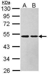
- Submitted by
- GeneTex (provider)
- Main image
- Experimental details
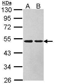
- Sample (30 ?g of whole cell lysate) A: HeLa B: HepG2 (GTX27900) 10% SDS PAGE GTX113629 diluted at 1:1000 The HRP-conjugated anti-rabbit IgG antibody (GTX213110-01) was used to detect the primary antibody.
Supportive validation
- Submitted by
- GeneTex (provider)
- Main image
- Experimental details
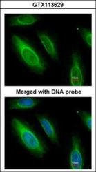
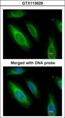
- Immunofluorescence analysis of paraformaldehyde-fixed HeLa, using Argininosuccinate Lyase(GTX113629) antibody at 1:200 dilution.
- Submitted by
- GeneTex (provider)
- Main image
- Experimental details
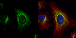
- ASL antibody detects ASL protein at cytoplasm by immunofluorescent analysis.Sample: HeLa cells were fixed in 4% paraformaldehyde at RT for 15 min.Green: ASL protein stained by ASL antibody (GTX113629) diluted at 1:500.Red: alpha Tubulin, a cytoskeleton marker, stained by alpha Tubulin antibody [GT114] (GTX628802) diluted at 1:1000.Blue: Hoechst 33342 staining.

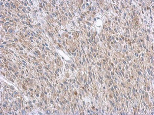
Supportive validation
- Submitted by
- GeneTex (provider)
- Main image
- Experimental details
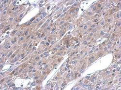
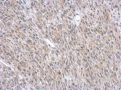
- Immunohistochemical analysis of paraffin-embedded U87 xenograft, using ASL(GTX113629) antibody at 1:500 dilution.
- Submitted by
- GeneTex (provider)
- Main image
- Experimental details
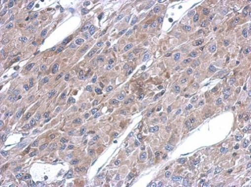
- Immunohistochemical analysis of paraffin-embedded C2C12 xenograft, using ASL(GTX113629) antibody at 1:500 dilution.