Explore
 Validate
 Learn
Learn Western blot
Antibody data
- Antibody Data
- Antigen structure
- References [5]
- Comments [0]
- Validations
- Western blot [2]
- Immunohistochemistry [1]
Submit
Validation data
Reference
Comment
Report error
- Product number
- MA5-12227 - Provider product page

- Provider
- Invitrogen Antibodies
- Product name
- Villin Monoclonal Antibody (CWWB1)
- Antibody type
- Monoclonal
- Antigen
- Other
- Description
- MA5-12227 targets Villin in IHC (P) applications and shows reactivity with Human samples. The MA5-12227 immunogen is human Villin protein.
- Reactivity
- Human, Mouse
- Host
- Mouse
- Isotype
- IgG
- Antibody clone number
- CWWB1
- Vial size
- 500 µL
- Concentration
- 0.2 mg/mL
- Storage
- 4° C
Submitted references NPC1L1-dependent transport of 27-alkyne cholesterol in intestinal epithelial cells.
Pulmonary enteric adenocarcinoma: a study of the clinicopathologic and molecular status of nine cases.
Oncogenic transformation of diverse gastrointestinal tissues in primary organoid culture.
Efficient differentiation of human embryonic stem cells to definitive endoderm.
Phenotype of columnar-lined esophagus in rats with esophagogastroduodenal anastomosis: similarity to human Barrett's esophagus.
Ticho AL, Calzadilla N, Malhotra P, Lee H, Anbazhagan AN, Saksena S, Dudeja PK, Lee D, Gill RK, Alrefai WA
American journal of physiology. Cell physiology 2021 May 1;320(5):C916-C925
American journal of physiology. Cell physiology 2021 May 1;320(5):C916-C925
Pulmonary enteric adenocarcinoma: a study of the clinicopathologic and molecular status of nine cases.
Wang CX, Liu B, Wang YF, Zhang RS, Yu B, Lu ZF, Shi QL, Zhou XJ
International journal of clinical and experimental pathology 2014;7(3):1266-74
International journal of clinical and experimental pathology 2014;7(3):1266-74
Oncogenic transformation of diverse gastrointestinal tissues in primary organoid culture.
Li X, Nadauld L, Ootani A, Corney DC, Pai RK, Gevaert O, Cantrell MA, Rack PG, Neal JT, Chan CW, Yeung T, Gong X, Yuan J, Wilhelmy J, Robine S, Attardi LD, Plevritis SK, Hung KE, Chen CZ, Ji HP, Kuo CJ
Nature medicine 2014 Jul;20(7):769-77
Nature medicine 2014 Jul;20(7):769-77
Efficient differentiation of human embryonic stem cells to definitive endoderm.
D'Amour KA, Agulnick AD, Eliazer S, Kelly OG, Kroon E, Baetge EE
Nature biotechnology 2005 Dec;23(12):1534-41
Nature biotechnology 2005 Dec;23(12):1534-41
Phenotype of columnar-lined esophagus in rats with esophagogastroduodenal anastomosis: similarity to human Barrett's esophagus.
Su Y, Chen X, Klein M, Fang M, Wang S, Yang CS, Goyal RK
Laboratory investigation; a journal of technical methods and pathology 2004 Jun;84(6):753-65
Laboratory investigation; a journal of technical methods and pathology 2004 Jun;84(6):753-65
No comments: Submit comment
Supportive validation
- Submitted by
- Invitrogen Antibodies (provider)
- Main image

- Experimental details
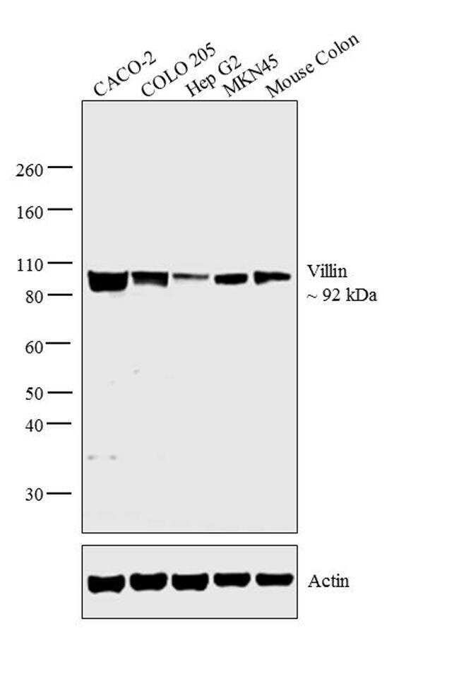
- Western blot analysis was performed on membrane-enriched extracts (30 µg lysate) of CACO-2 (Lane 1), COLO 205 (Lane 2), Hep G2 (Lane 3), MKN45 (Lane 4), and tissue extract (30 µg lysate) of mouse colon (Lane 5). The blots were probed with Mouse Anti-Villin Monoclonal Antibody (Product # MA5-12227, 2 µg/mL) and detected by chemiluminescence using Goat anti-Mouse IgG (H+L) Superclonal™ Secondary Antibody, HRP conjugate (Product # A28177, 0.25 µg/mL, 1:4000 dilution). A 92 kDa band corresponding to Villin was observed across the cell lines and tissues tested. Known quantity of protein samples were electrophoresed using Novex® NuPAGE® 4-12 % Bis-Tris gel (Product # NP0321BOX), XCell SureLock™ Electrophoresis System (Product # EI0002) and Novex® Sharp Pre-Stained Protein Standard (Product # LC5800). Resolved proteins were then transferred onto a nitrocellulose membrane with Pierce™ Power Blotter (Product # 22834). The membrane was probed with the relevant primary and secondary Antibody following blocking with 5 % skimmed milk. Chemiluminescent detection was performed using Pierce™ ECL Western Blotting Substrate (Product # 32106).
- Submitted by
- Invitrogen Antibodies (provider)
- Main image
- Experimental details
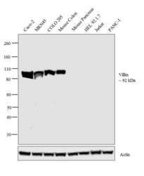
- Western blot analysis was performed on membrane enriched extracts (30 µg lysate) of Caco-2 (Lane 1), MKN45 (Lane 2), COLO 205 (Lane 3), HEL 92.1.7 (Lane 6), Jurkat (Lane 7), PANC-1 (Lane 8) and tissue extracts (30 µg lysate) of Mouse Colon (Lane 4) and Mouse Pancreas (Lane 5). The blot was probed with anti-Villin Mouse Monoclonal Antibody (Product # MA5-12227, 2 µg/mL) and detected by chemiluminescence using Goat anti-Mouse IgG (H+L) Superclonal™ Secondary Antibody, HRP conjugate (Product # A28177, 0.25 µg/mL, 1:4000 dilution). A 92 kDa band corresponding to Villin was observed in Caco-2, MKN45, COLO 205, Mouse Colon and not observed in other cell lines and tissues which are documented to be Villin negative.

Supportive validation
- Submitted by
- Invitrogen Antibodies (provider)
- Main image

- Experimental details
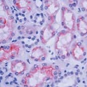
- Formalin-fixed, paraffin-embedded human kidney stained with Villin antibody using peroxidase-conjugate and AEC chromogen. Note brush border and cytoplasmic staining of epithelial cells in the proximal tubules.