 Explore
 Validate
 Learn
Learn Western blot
 ELISA
ELISAAntibody data
- Antibody Data
- Antigen structure
- References [16]
- Comments [0]
- Validations
- Western blot [1]
- Immunocytochemistry [1]
- Immunoprecipitation [1]
- Immunohistochemistry [2]
Submit
Validation data
Reference
Comment
Report error
- Product number
- 66096-1-Ig - Provider product page

- Provider
- Proteintech Group
- Product name
- Villin antibody
- Antibody type
- Monoclonal
- Description
- Villin antibody (Cat. #66096-1-Ig) is a mouse monoclonal antibody that shows reactivity with human, mouse and has been validated for the following applications: IF, IHC, IP, WB,ELISA.
- Reactivity
- Human, Mouse
- Host
- Mouse
- Conjugate
- Unconjugated
- Isotype
- IgG
- Antibody clone number
- 2B7B9
- Vial size
- 20ul, 150ul
Submitted references 4-Octyl itaconate attenuates radiation-induced intestinal injury associated with ferroptosis inhibition and microbiota rebalance.
L-theanine's therapeutic effects on colorectal cancer: Mechanistic insights from a 1,2-dimethylhydrazine-induced rat model.
Bupleuri Radix Polysaccharides Alleviate MASLD by Regulating Muribaculaceae-Derived SCFAs in the Gut-Liver Axis.
Serum Villin-1-A Novel Marker of Gut Barrier Damage in Acutely Decompensated Cirrhosis: A Cohort Study and Validation.
Sinisan, a compound Chinese herbal medicine, alleviates acute colitis by facilitating colonic secretory cell lineage commitment and mucin production.
Hepatic FXR-FGF4 is required for bile acid homeostasis via an FGFR4-LRH-1 signal node under cholestatic stress.
Development of Sheep Intestinal Organoids for Studying Deoxynivalenol-Induced Toxicity.
Excessive Mitochondrial Fission Suppresses Mucosal Repair by Impairing Butyrate Metabolism in Colonocytes.
Methylthioacetic acid, a derivative of aroma compounds from Cucumis melo var. conomon dose-dependently triggers differentiation and apoptosis of RCM-1 human colorectal cancer cells.
Stiffness Restricts the Stemness of the Intestinal Stem Cells and Skews Their Differentiation Toward Goblet Cells.
Protective effect of total flavonoids of Engelhardia roxburghiana Wall. leaves against radiation-induced intestinal injury in mice and its mechanism.
Sitagliptin Alleviates Radiation-Induced Intestinal Injury by Activating NRF2-Antioxidant Axis, Mitigating NLRP3 Inf--lammasome Activation, and Reversing Gut Microbiota Disorder.
Syntaxin 3 interacts with serotonin transporter and regulates its function.
The Circadian Clock Gene Bmal1 Controls Intestinal Exporter MRP2 and Drug Disposition.
A three-protein signature and clinical outcome in esophageal squamous cell carcinoma.
A four actin-binding protein signature model for poor prognosis of patients with esophageal squamous cell carcinoma.
Zhang S, Yue T, Jin P, Zhang X, Huo Q, Li W, Tian C, Dong H, Dong Y, Zhao Y, Li D
Free radical biology & medicine 2026 Jul;250:386-398
Free radical biology & medicine 2026 Jul;250:386-398
L-theanine's therapeutic effects on colorectal cancer: Mechanistic insights from a 1,2-dimethylhydrazine-induced rat model.
Jimusi, Han Y, Su X, Li Q, Liu S, Cheng S, Shen W, He J, Yuesitu, Nashunbayaer
Translational oncology 2026 Jan;63:102609
Translational oncology 2026 Jan;63:102609
Bupleuri Radix Polysaccharides Alleviate MASLD by Regulating Muribaculaceae-Derived SCFAs in the Gut-Liver Axis.
Yang Y, Wang H, Gu Y, Wu R, Qin W, Chen R, Fan G, Xue X, Lan J, Huang Z, Han Q, Liu R
International journal of molecular sciences 2026 Jan 8;27(2)
International journal of molecular sciences 2026 Jan 8;27(2)
Serum Villin-1-A Novel Marker of Gut Barrier Damage in Acutely Decompensated Cirrhosis: A Cohort Study and Validation.
Tornai D, Balogh B, Csillag A, Budai A, Kiss A, Antal-Szalmas P, Mehes G, Barath L, Tornai T, Tornai I, Vitalis Z, Sipeki N, Dinya T, Enyedi A, Rosenberger FA, Laleman W, Coenraad MJ, Aguilar F, Claria J, Moreau R, Trebicka J, Papp M, MICROB‐PREDICT and PREDICT study group of the EASL‐CLIF consortium
Alimentary pharmacology & therapeutics 2026 Apr;63(7):1018-1032
Alimentary pharmacology & therapeutics 2026 Apr;63(7):1018-1032
Sinisan, a compound Chinese herbal medicine, alleviates acute colitis by facilitating colonic secretory cell lineage commitment and mucin production.
Cai YJ, Lan JH, Li S, Feng YN, Li FH, Guo MY, Liu RP
Journal of integrative medicine 2025 Jul;23(4):429-444
Journal of integrative medicine 2025 Jul;23(4):429-444
Hepatic FXR-FGF4 is required for bile acid homeostasis via an FGFR4-LRH-1 signal node under cholestatic stress.
Song L, Hou Y, Xu D, Dai X, Luo J, Liu Y, Huang Z, Yang M, Chen J, Hu Y, Chen C, Tang Y, Rao Z, Ma J, Zheng M, Shi K, Cai C, Lu M, Tang R, Ma X, Xie C, Luo Y, Li X, Huang Z
Cell metabolism 2025 Jan 7;37(1):104-120.e9
Cell metabolism 2025 Jan 7;37(1):104-120.e9
Development of Sheep Intestinal Organoids for Studying Deoxynivalenol-Induced Toxicity.
Wang H, He X, Zhang M, Fan N, Yang Z, Shen T, Guo J, Song Y, Cao G, Liu Y, Li X, Nashun B
International journal of molecular sciences 2025 Jan 23;26(3)
International journal of molecular sciences 2025 Jan 23;26(3)
Excessive Mitochondrial Fission Suppresses Mucosal Repair by Impairing Butyrate Metabolism in Colonocytes.
Fu SC, Qu JY, Li LX, Yang XX, Li YQ, Zuo XL
Inflammatory bowel diseases 2024 Jan 5;30(1):114-124
Inflammatory bowel diseases 2024 Jan 5;30(1):114-124
Methylthioacetic acid, a derivative of aroma compounds from Cucumis melo var. conomon dose-dependently triggers differentiation and apoptosis of RCM-1 human colorectal cancer cells.
Kamimura M, Sasaki A, Otani Y, Nakamura Y, Nakamura T, Kuramochi K, Imai T, Kubo N, Okamoto S
The Journal of toxicological sciences 2023;48(1):25-35
The Journal of toxicological sciences 2023;48(1):25-35
Stiffness Restricts the Stemness of the Intestinal Stem Cells and Skews Their Differentiation Toward Goblet Cells.
He S, Lei P, Kang W, Cheung P, Xu T, Mana M, Park CY, Wang H, Imada S, Russell JO, Wang J, Wang R, Zhou Z, Chetal K, Stas E, Mohad V, Bruun-Rasmussen P, Sadreyev RI, Hodin RA, Zhang Y, Breault DT, Camargo FD, Yilmaz ÖH, Fredberg JJ, Saeidi N
Gastroenterology 2023 Jun;164(7):1137-1151.e15
Gastroenterology 2023 Jun;164(7):1137-1151.e15
Protective effect of total flavonoids of Engelhardia roxburghiana Wall. leaves against radiation-induced intestinal injury in mice and its mechanism.
Wu S, Tian C, Tu Z, Guo J, Xu F, Qin W, Chang H, Wang Z, Hu T, Sun X, Ning H, Li Y, Gou W, Hou W
Journal of ethnopharmacology 2023 Jul 15;311:116428
Journal of ethnopharmacology 2023 Jul 15;311:116428
Sitagliptin Alleviates Radiation-Induced Intestinal Injury by Activating NRF2-Antioxidant Axis, Mitigating NLRP3 Inf--lammasome Activation, and Reversing Gut Microbiota Disorder.
Huang S, Huang Y, Lin W, Wang L, Yang Y, Li P, Xiao L, Chen Y, Chu Q, Yuan X
Oxidative medicine and cellular longevity 2022;2022:2586305
Oxidative medicine and cellular longevity 2022;2022:2586305
Syntaxin 3 interacts with serotonin transporter and regulates its function.
Motoike S, Taguchi K, Harada K, Asano M, Hide I, Tanaka S, Irifune M, Sakai N
Journal of pharmacological sciences 2021 Apr;145(4):297-307
Journal of pharmacological sciences 2021 Apr;145(4):297-307
The Circadian Clock Gene Bmal1 Controls Intestinal Exporter MRP2 and Drug Disposition.
Yu F, Zhang T, Zhou C, Xu H, Guo L, Chen M, Wu B
Theranostics 2019;9(10):2754-2767
Theranostics 2019;9(10):2754-2767
A three-protein signature and clinical outcome in esophageal squamous cell carcinoma.
Cao HH, Zhang SY, Shen JH, Wu ZY, Wu JY, Wang SH, Li EM, Xu LY
Oncotarget 2015 Mar 10;6(7):5435-48
Oncotarget 2015 Mar 10;6(7):5435-48
A four actin-binding protein signature model for poor prognosis of patients with esophageal squamous cell carcinoma.
Peng ZM, Yu W, Xie Y, Peng WH, Cao HH, Shen JH, Wu ZY, Li EM, Xu LY
International journal of clinical and experimental pathology 2014;7(9):5950-9
International journal of clinical and experimental pathology 2014;7(9):5950-9
No comments: Submit comment
Supportive validation
- Submitted by
- Proteintech Group (provider)
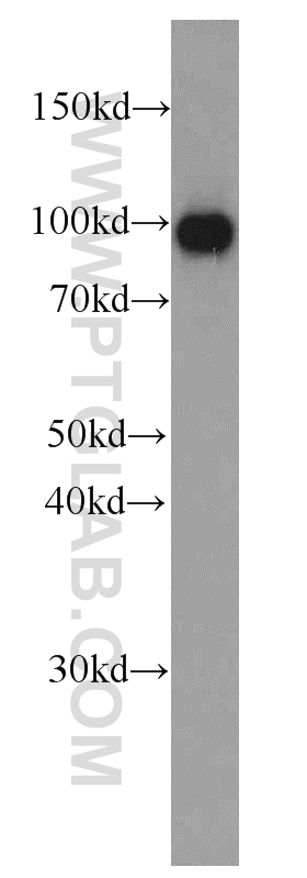
- Main image

- Experimental details
- human kidney tissue were subjected to SDS PAGE followed by western blot with 66096-1-Ig(VIL1 antibody) at dilution of 1:1000
- Sample type
- tissue
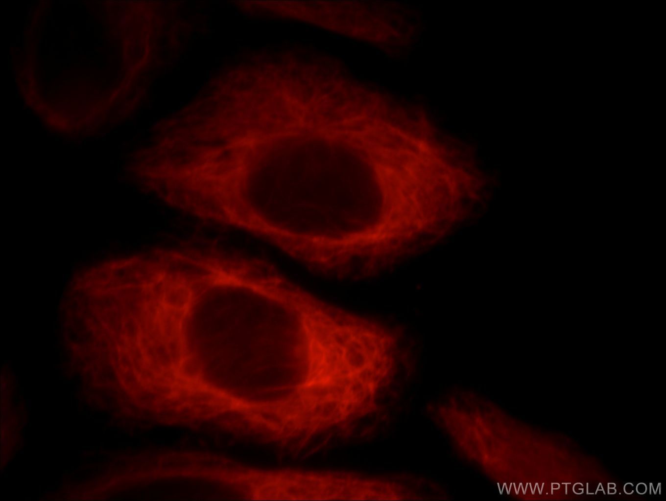
Supportive validation
- Submitted by
- Proteintech Group (provider)
- Main image

- Experimental details
- Immunofluorescent analysis of HepG2 cells, using VIL1 antibody 66096-1-lg at 1:25 dilution and Rhodamine-labeled goat anti-mouse IgG (red).
- Sample type
- cell line
Supportive validation
- Submitted by
- Proteintech Group (provider)
- Main image
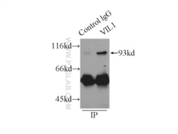
- Experimental details
- IP Result of anti-VIL1 (IP:66096-1-Ig, 4ug; Detection:66096-1-Ig 1:1000) with mouse kidney tissue lysate 6000ug.
- Sample type
- tissue
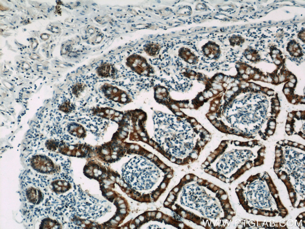
Supportive validation
- Submitted by
- Proteintech Group (provider)
- Main image

- Experimental details
- Immunohistochemical of paraffin-embedded human small intestine using 66096-1-Ig(VIL1 antibody) at dilution of 1:500 (under 10x lens)
- Sample type
- tissue
- Submitted by
- Proteintech Group (provider)
- Main image
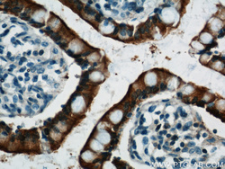
- Experimental details
- Immunohistochemical of paraffin-embedded human small intestine using 66096-1-Ig(VIL1 antibody) at dilution of 1:500 (under 40x lens)
- Sample type
- tissue
