Explore
 Validate
 Learn
Learn Western blot
 Immunocytochemistry
Antibody data
- Antibody Data
- Antigen structure
- References [3]
- Comments [0]
- Validations
- Immunocytochemistry [1]
Submit
Validation data
Reference
Comment
Report error
- Product number
- HPA006884 - Provider product page

- Provider
- Atlas Antibodies
- Proper citation
- Atlas Antibodies Cat#HPA006884, RRID:AB_1080566
- Product name
- Anti-VIL1
- Antibody type
- Polyclonal
- Description
- Polyclonal Antibody against Human VIL1, Gene description: villin 1, Alternative Gene Names: D2S1471, VIL, Validated applications: IHC, WB, ICC, Uniprot ID: P09327, Storage: Store at +4°C for short term storage. Long time storage is recommended at -20°C.
- Reactivity
- Human
- Host
- Rabbit
- Conjugate
- Unconjugated
- Isotype
- IgG
- Vial size
- 100 µl
- Concentration
- 0.3 mg/ml
- Storage
- Store at +4°C for short term storage. Long time storage is recommended at -20°C.
- Handling
- The antibody solution should be gently mixed before use.
Submitted references Villin is a biomarker for reverse polarity in colorectal micropapillary carcinoma
Antibodies Biotinylated Using a Synthetic Z-domain from Protein A Provide Stringent In Situ Protein Detection
Antibodies biotinylated using a synthetic Z-domain from protein A provide stringent in situ protein detection.
Zhao L, Liu S, Li Y, Xiong Z
Oncology Letters 2020;21(1)
Oncology Letters 2020;21(1)
Antibodies Biotinylated Using a Synthetic Z-domain from Protein A Provide Stringent In Situ Protein Detection
Andersson S, Konrad A, Ashok N, Ponten F, Hober S, Asplund A
Journal of Histochemistry & Cytochemistry 2013 October;61(11):773-784
Journal of Histochemistry & Cytochemistry 2013 October;61(11):773-784
Antibodies biotinylated using a synthetic Z-domain from protein A provide stringent in situ protein detection.
Andersson S, Konrad A, Ashok N, Pontén F, Hober S, Asplund A
The journal of histochemistry and cytochemistry : official journal of the Histochemistry Society 2013 Nov;61(11):773-84
The journal of histochemistry and cytochemistry : official journal of the Histochemistry Society 2013 Nov;61(11):773-84
No comments: Submit comment
Supportive validation
- Submitted by
- Atlas Antibodies (provider)
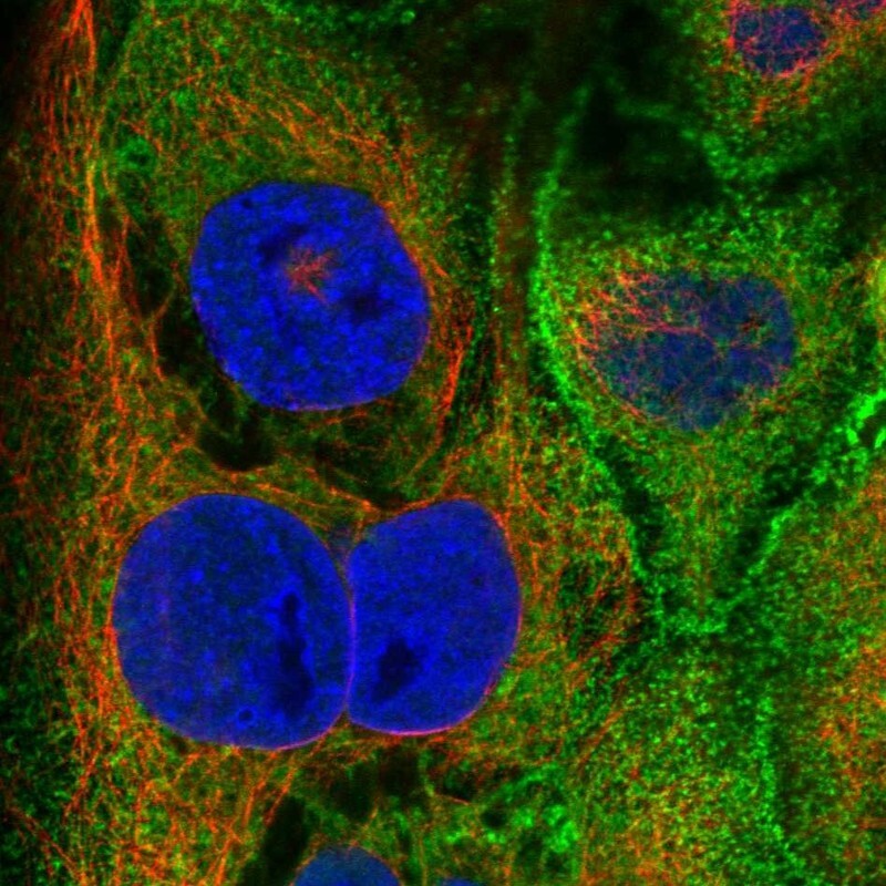
- Main image

- Experimental details
- Immunofluorescent staining of human cell line CACO-2 shows localization to plasma membrane.
- Sample type
- Human