 Explore
 Validate
 Learn
LearnABIN2509152
antibody from antibodies-online
Targeting: RPS6KB2
KLS, P70-BETA, p70S6Kb, S6KB, S6Kbeta, S6Kβ, STK14B
 Western blot
Antibody data
- Antibody Data
- Antigen structure
- References [0]
- Comments [0]
- Validations
- Western blot [1]
- Immunocytochemistry [3]
- Immunohistochemistry [2]
Submit
Validation data
Reference
Comment
Report error
- Product number
- ABIN2509152 - Provider product page

- Provider
- antibodies-online
- Product name
- anti-Ribosomal Protein S6 Kinase, 70kDa, Polypeptide 2 (RPS6KB2) antibody
- Antibody type
- Monoclonal
- Antigen
- This RPS6KB2 monoclonal antibody is generated from mouse immunized with RPS6KB2 recombinant protein.Antigen type: Recombinant Protein
- Description
- This antibody is purified through a protein G column, followed by dialysis against PBS.
- Reactivity
- Human, Mouse
- Host
- Mouse
- Isotype
- IgG
- Vial size
- 200 μL
- Storage
- Maintain refrigerated at 2-8°C for up to 6 months. For long term storage store at -20°C in small aliquots to prevent freeze-thaw cycles.
No comments: Submit comment
Supportive validation
- Submitted by
- antibodies-online (provider)
- Main image
- Experimental details
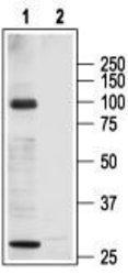
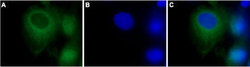
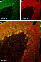
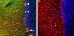
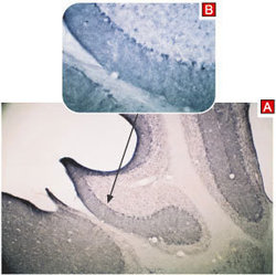
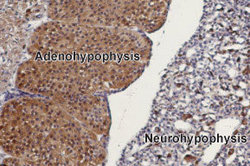
- Western blot analysis of rat heart membranes: 1. Anti-TRPC3 antibody (ABIN2511203), (1:200). 2. Anti-TRPC3 antibody, preincubated with the control peptide antigen. Expression of TRPC3 in rat pituitary gland Immunohistochemical staining of rat pituitary gland paraffin embedded sections using Anti-TRPC3 antibody (ABIN2511203), (1:100). TRPC3 is mainly expressed in the adenohypophysis (on left). Hematoxilin is used as the counterstain. Expression of TRPC3 in mouse cerebellum Immunohistochemical staining of mouse cerebellum using Anti-TRPC3 antibody (ABIN2511203) (A). Immunoreactivity appears in the molecular layer and in Purkinje cells (B). Expression of TRPC3 in rat cerebellum Immunohistochemical staining of TRPC3 in rat cerebellum using Anti-TRPC3 antibody (ABIN2511203). A. TRPC3 (green) appears in Purkinje cells (arrows) including both soma and dendrites and as well as in the molecular layer neuropil (asterisk). Staining of the same section with mouse anti-parvalbumin (red) reveals that TRPC3 is not expressed in molecular layer interneurons. B. Pre-incubating Anti-TRPC3 with the TRPC3 peptide antigen blocks staining. DAPI is used as the counterstain (blue). Colocalization of TRPC6 and TRPC3 in rat cerebellum Immunohistochemical staining of rat cerebellum frozen section using guinea pig Anti-TRPC6 antibody (AGP-002) and rabbit Anti-TRPC3 antibody (ABIN2511203). A. TRPC6 staining (green) appears in molecular layer and in Purkinje cells. B. In the same section, staining of TRPC3 (red) appears as well in both molecular layer and Purkinje cells. C. Merge images of A and B indicates extensive co-localization. DAPI is used as the counterstain (blue). Expression of TRPC3 in rat C6 brain glioma cells Immunocytochemical staining of Paraformaldehyde-fixed and permeabilized rat C6 brain glioma cells. A. Staining using Anti-TRPC3 antibody (ABIN2511203), (1:500) followed by goat anti-rabbit-AlexaFluor-488 secondary antibody. B. Nuclear staining using the cell-permeable dye Hoechst 33342. C. Merged image of panels A and B.

Supportive validation
- Submitted by
- antibodies-online (provider)
- Main image
- Experimental details
- Image(s): Immunofluorescence

- Submitted by
- antibodies-online (provider)
- Main image
- Experimental details
- Image(s): Immunofluorescence

- Submitted by
- antibodies-online (provider)
- Main image
- Experimental details
- Image(s): Immunofluorescence

Supportive validation
- Submitted by
- antibodies-online (provider)
- Main image
- Experimental details
- Image(s): Immunohistochemistry

- Submitted by
- antibodies-online (provider)
- Main image
- Experimental details
- Image(s): Immunohistochemistry
