Explore
 Validate
 Learn
Learn Western blot
 Immunohistochemistry
Antibody data
- Antibody Data
- Antigen structure
- References [2]
- Comments [0]
- Validations
- Immunohistochemistry [2]
Submit
Validation data
Reference
Comment
Report error
- Product number
- AF1028 - Provider product page

- Provider
- Novus Biologicals
- Product name
- Goat Polyclonal CD30 Ligand/TNFSF8 Antibody
- Antibody type
- Polyclonal
- Description
- Antigen Affinity-purified. Detects human and mouse CD30 Ligand in direct ELISAs and Western blots. In direct ELISAs, less than 10% cross-reactivity with recombinant human (rh) CD27 Ligand is observed and less than 1% cross-reactivity with rhCD40 Ligand is observed.
- Reactivity
- Human
- Host
- Goat
- Conjugate
- Unconjugated
- Isotype
- IgG
- Vial size
- 100 ug
- Concentration
- LYOPH
- Storage
- Use a manual defrost freezer and avoid repeated freeze-thaw cycles. 12 months from date of receipt, -20 to -70 degreesC as supplied. 1 month, 2 to 8 degreesC under sterile conditions after reconstitution. 6 months, -20 to -70 degreesC under sterile conditions after reconstitution.
Submitted references Selective redox regulation of cytokine receptor signaling by extracellular thioredoxin-1.
CD40 induces macrophage anti-Toxoplasma gondii activity by triggering autophagy-dependent fusion of pathogen-containing vacuoles and lysosomes.
Schwertassek U, Balmer Y, Gutscher M, Weingarten L, Preuss M, Engelhard J, Winkler M, Dick TP
The EMBO journal 2007 Jul 11;26(13):3086-97
The EMBO journal 2007 Jul 11;26(13):3086-97
CD40 induces macrophage anti-Toxoplasma gondii activity by triggering autophagy-dependent fusion of pathogen-containing vacuoles and lysosomes.
Andrade RM, Wessendarp M, Gubbels MJ, Striepen B, Subauste CS
The Journal of clinical investigation 2006 Sep;116(9):2366-77
The Journal of clinical investigation 2006 Sep;116(9):2366-77
No comments: Submit comment
Supportive validation
- Submitted by
- Novus Biologicals (provider)
- Main image

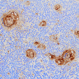
- Experimental details
- CD30 Ligand/TNFSF8 in Human Thymus. CD30 Ligand/TNFSF8 was detected in immersion fixed paraffin-embedded sections of human thymus using Goat Anti-Human CD30 Ligand/TNFSF8 Antigen Affinity-purified Polyclonal Antibody (Catalog # AF1028) at 0.3 µg/mL for 1 hour at room temperature followed by incubation with the Anti-Goat IgG VisUCyte™ HRP Polymer Antibody (Catalog # VC004). Tissue was stained using DAB (brown) and counterstained with hematoxylin (blue). Specific staining was localized to Hassall's corpuscles. View our protocol for IHC Staining with VisUCyte HRP Polymer Detection Reagents.
- Submitted by
- Novus Biologicals (provider)
- Main image

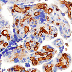
- Experimental details
- CD30 Ligand/TNFSF8 in Human Placenta. CD30 Ligand/TNFSF8 was detected in immersion fixed paraffin-embedded sections of human placenta using Goat Anti-Human CD30 Ligand/TNFSF8 Antigen Affinity-purified Polyclonal Antibody (Catalog # AF1028) at 0.1 µg/mL for 1 hour at room temperature followed by incubation with the Anti-Goat IgG VisUCyte™ HRP Polymer Antibody (Catalog # VC004). Tissue was stained using DAB (brown) and counterstained with hematoxylin (blue). Specific staining was localized to endothelial cells in villi. View our protocol for IHC Staining with VisUCyte HRP Polymer Detection Reagents.