Explore
 Validate
 Learn
Learn Western blot
 Immunoprecipitation
Antibody data
- Antibody Data
- Antigen structure
- References [0]
- Comments [0]
- Validations
- Western blot [3]
- Immunocytochemistry [2]
Submit
Validation data
Reference
Comment
Report error
- Product number
- PA5-19584 - Provider product page

- Provider
- Invitrogen Antibodies
- Product name
- Menin Polyclonal Antibody
- Antibody type
- Polyclonal
- Antigen
- Synthetic peptide
- Description
- This antibody is predicted to react with cow based on sequence homology.
- Reactivity
- Human, Mouse, Rat
- Host
- Rabbit
- Isotype
- IgG
- Vial size
- 200 µL
- Concentration
- 0.5 mg/mL
- Storage
- Store at 4°C short term. For long term storage, store at -20°C, avoiding freeze/thaw cycles.
No comments: Submit comment
Supportive validation
- Submitted by
- Invitrogen Antibodies (provider)
- Main image
- Experimental details
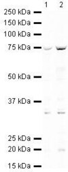
- Western blot analysis of HeLa Whole Cell Lysate using Product # PA5-19584, Menin primary antibody at a dilution of 1 µg/mL (lane 1). Staining of Jurkat Whole Cell Lysate at a dilution of 1 µg/mL (lane 2). Blot treated with a secondary IR Dye680-conjugated Goat polyclonal anti-Rabbit antibody was used at a dilution of 1:10000.

- Submitted by
- Invitrogen Antibodies (provider)
- Main image
- Experimental details
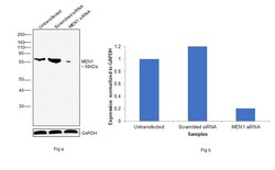
- Knockdown of MEN1 was achieved by transfecting HEK 293 with MEN1 specific (Silencer® select Product # s8684). Western blot analysis (Fig. a) was performed using modified whole cell extracts (1% SDS) from the untransfected cells (Lane 1), non-specific scrambled siRNA transfected cells (Lane 2) and MEN1 knockdown cells (Lane 3). The blot was probed with MEN1 Polyclonal Antibody(Product # PA5-19584, 1 µg/mL) and Goat anti-Rabbit IgG (H+L) Superclonal™ Recombinant Secondary Antibody, HRP (Product # A27036, 1:4000 dilution). Densitometric analysis of this western blot is shown i histogram (Fig. b). Loss of signal upon siRNA mediated knock down confirms that antibody is specific to MEN1.
- Submitted by
- Invitrogen Antibodies (provider)
- Main image

- Experimental details
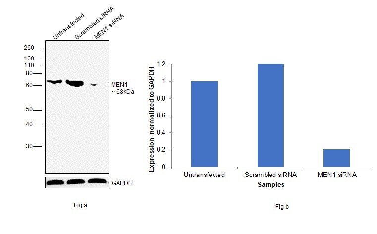
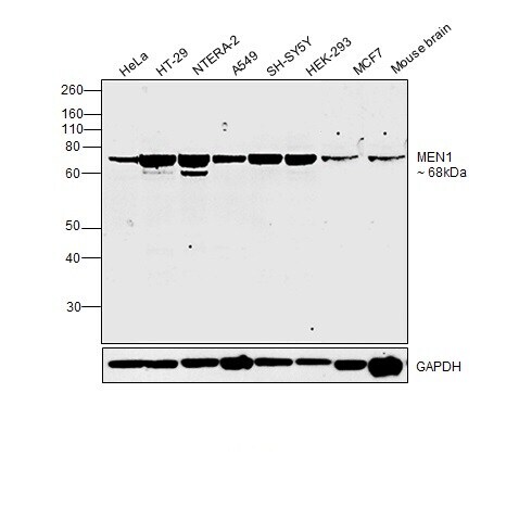
- Western blot was performed using Menin Polyclonal Antibody (Product # PA5-19584) and a 68 kDa band corresponding to MEN1 was observed across cell lines and tissues tested. Whole cell extracts (1% SDS) (30 µg lysate) of HeLa (Lane 1), HT-29 (Lane 2), NTERA-2 (Lane 3), A549 (Lane 4), SH SY5Y (Lane 5), HEK 293 (Lane 6), MCF7 (Lane 7) and Mouse brain (Lane 8) were electrophoresed using NuPAGE® 4-12 % Bis-Tris gel (Product # NP0322BOX)Resolved proteins were then transferred onto a nitrocellulose membrane (Product # IB23001) by iBlot® 2 Dry Blotting System (Product # IB21001). The blot was probed with the primary antibody (1 µg/mL) and detected by chemiluminescence with Goat anti-Rabbit IgG (H+L), Superclonal™ Recombinant Secondary Antibody, HRP (Product # A27036, 1:4000 dilution) using the iBright FL 1000 (Product # A32752). Chemiluminescent detection was performed using Novex® ECL Chemiluminescent Substrate Reagent Kit (Product # WP20005).
Supportive validation
- Submitted by
- Invitrogen Antibodies (provider)
- Main image

- Experimental details
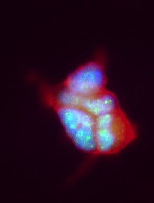
- Immunofluorescent staining of HEK 293 using Product # PA5-19584, anti-Menin antibody. The cells were fixed with PFA (4%) for 10 minutes, permabilised with BSA (1%), normal goat serum (10%) and glycine (0.3 M) in 0.1% T-BST for 20 minutes and exposed to the primary antibody at a concentration of 1 µg/mL for 1 hour at room temp. The secondary antibody was a 448 fluorescence conjugated Goat anti-rabbit IgG (green) at a dilution of 1:1000. A WGA- 594 fluorescent conjugated stain was used to label plasma membranes (red) and the nuclei stain was DAPI (blue).

- Submitted by
- Invitrogen Antibodies (provider)
- Main image

- Experimental details
- Immunofluorescent staining of HEK 293 using Product # PA5-19584, anti-Menin antibody. The cells were fixed with PFA (4%) for 10 minutes, permabilised with BSA (1%), normal goat serum (10%) and glycine (0.3 M) in 0.1% T-BST for 20 minutes and exposed to the primary antibody at a concentration of 1 µg/mL for 1 hour at room temp. The secondary antibody was a 448 fluorescence conjugated Goat anti-rabbit IgG (green) at a dilution of 1:1000. A WGA- 594 fluorescent conjugated stain was used to label plasma membranes (red) and the nuclei stain was DAPI (blue).