Explore
 Validate
 Learn
Learn Western blot
 Immunocytochemistry
Antibody data
- Antibody Data
- Antigen structure
- References [0]
- Comments [0]
- Validations
- Immunocytochemistry [1]
- Immunohistochemistry [6]
Submit
Validation data
Reference
Comment
Report error
- Product number
- PA5-84020 - Provider product page

- Provider
- Invitrogen Antibodies
- Product name
- PIT1 Polyclonal Antibody
- Antibody type
- Polyclonal
- Antigen
- Recombinant protein fragment
- Description
- Immunogen sequence: FPDHTLSHGF PPIHQPLLAE DPTAADFKQE LRRKSKLVEE PIDMDSPEIR ELEKFANEFK VRRIKLGYTQ TNVGEALAAV HGS
- Reactivity
- Human
- Host
- Rabbit
- Isotype
- IgG
- Vial size
- 100 μL
- Concentration
- 0.2 mg/mL
- Storage
- Store at 4°C short term. For long term storage, store at -20°C, avoiding freeze/thaw cycles.
No comments: Submit comment
Supportive validation
- Submitted by
- Invitrogen Antibodies (provider)
- Main image
- Experimental details
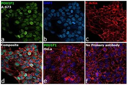
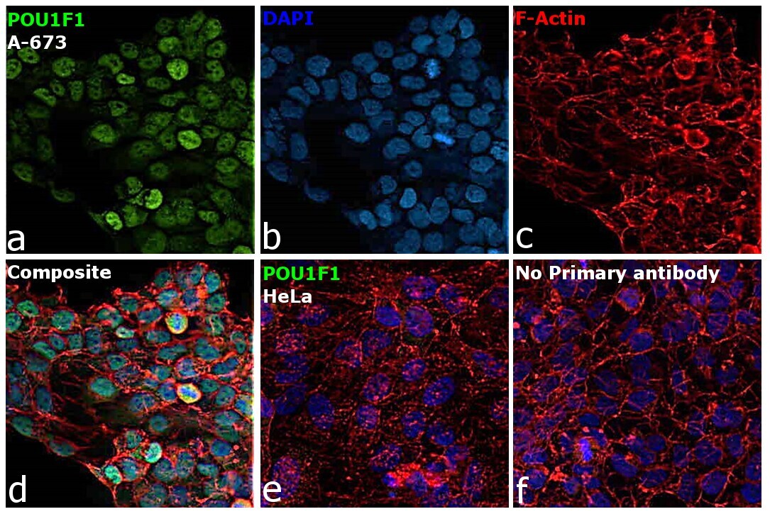
- Immunofluorescence analysis of PIT1/POU1F1 was performed using 70% confluent log phase A-673 and HeLa cells. The cells were fixed with 4% paraformaldehyde for 10 minutes, permeabilized with 0.1% Triton™ X-100 for 15 minutes, and blocked with 2% BSA for 1 hour at room temperature. The cells were labeled with PIT1 Polyclonal Antibody (Product # PA5-84020, 1:100 dilution) in 0.1% BSA, incubated at 4 degree celsius overnight and then labeled with Donkey anti-Rabbit IgG (H+L) Highly Cross-Adsorbed Secondary Antibody, Alexa Fluor™ Plus 488 (Product # A32790, 1:2000), for 45 minutes at room temperature (Panel a: Green). Nuclei (Panel b:Blue) were stained with ProLong™ Diamond Antifade Mountant with DAPI (Product # P36962). F-actin (Panel c: Red) was stained with Rhodamine Phalloidin (Product # R415, 1:300). Panel d represents the merged image showing nuclear localization of POU1F1/PIT1 in A-673 cell line. Panel e represents the merged image showing no signal in HeLa cell line. Panel f represents control A-673 cells with no primary antibody to assess background. The images were captured at 40X magnification.
Supportive validation
- Submitted by
- Invitrogen Antibodies (provider)
- Main image
- Experimental details
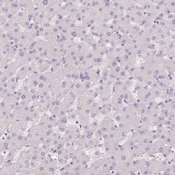
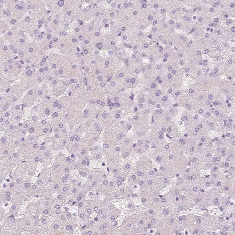
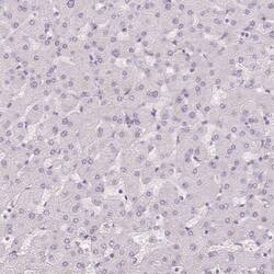
- Immunohistochemical analysis of PIT1 in human liver using PIT1 Polyclonal Antibody (Product # PA5-84020) shows no positivity in hepatocytes as expected.
- Submitted by
- Invitrogen Antibodies (provider)
- Main image
- Experimental details
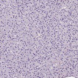
- Immunohistochemical analysis of PIT1 in human pancreas using PIT1 Polyclonal Antibody (Product # PA5-84020) shows no positivity in exocrine glandular cells as expected.

- Submitted by
- Invitrogen Antibodies (provider)
- Main image
- Experimental details
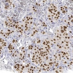
- Immunohistochemical analysis of PIT1 in human pituitary gland using PIT1 Polyclonal Antibody (Product # PA5-84020) shows moderate to strong nuclear positivity in neuroendocrine cells in the anterior lobe.

- Submitted by
- Invitrogen Antibodies (provider)
- Main image
- Experimental details
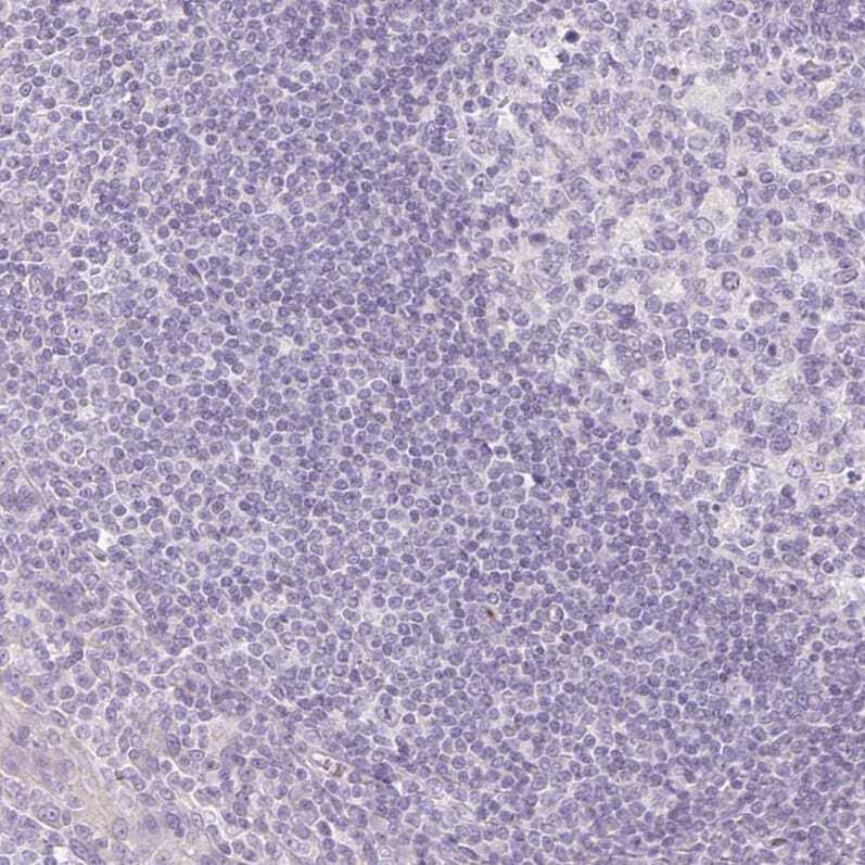
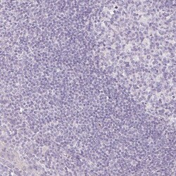
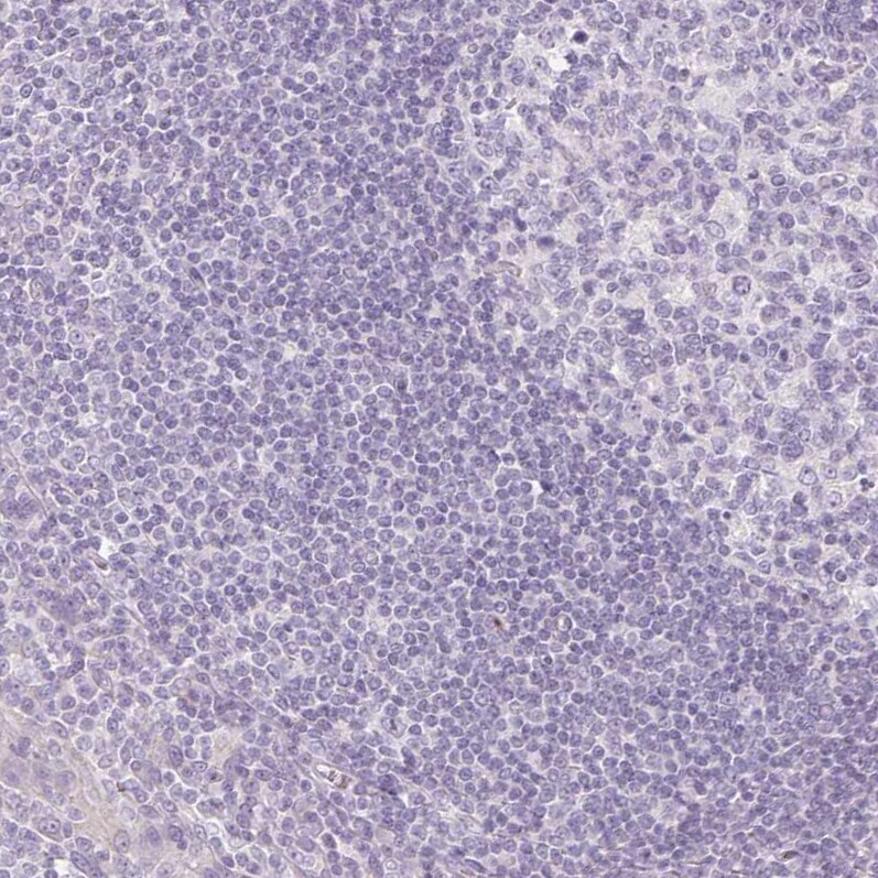
- Immunohistochemical analysis of PIT1 in human tonsil using PIT1 Polyclonal Antibody (Product # PA5-84020) shows no positivity in non-germinal center cells as expected.
- Submitted by
- Invitrogen Antibodies (provider)
- Main image
- Experimental details
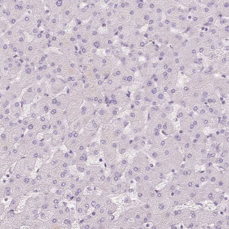
- Immunohistochemical analysis of PIT1 in human liver using PIT1 Polyclonal Antibody (Product # PA5-84020) shows no positivity in hepatocytes as expected.
- Submitted by
- Invitrogen Antibodies (provider)
- Main image
- Experimental details
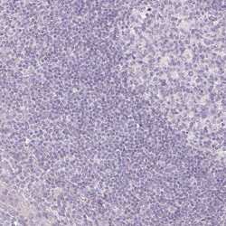
- Immunohistochemical analysis of PIT1 in human tonsil using PIT1 Polyclonal Antibody (Product # PA5-84020) shows no positivity in non-germinal center cells as expected.