Explore
 Validate
 Learn
Learn Western blot
 Immunocytochemistry
Antibody data
- Antibody Data
- Antigen structure
- References [1]
- Comments [0]
- Validations
- Immunocytochemistry [1]
- Immunohistochemistry [1]
Submit
Validation data
Reference
Comment
Report error
- Product number
- HPA024305 - Provider product page

- Provider
- Atlas Antibodies
- Proper citation
- Atlas Antibodies Cat#HPA024305, RRID:AB_1849922
- Product name
- Anti-GPI
- Antibody type
- Polyclonal
- Description
- Polyclonal Antibody against Human GPI, Gene description: glucose-6-phosphate isomerase, Alternative Gene Names: AMF, NLK, Validated applications: ICC, IHC, WB, Uniprot ID: P06744, Storage: Store at +4°C for short term storage. Long time storage is recommended at -20°C.
- Reactivity
- Human
- Host
- Rabbit
- Conjugate
- Unconjugated
- Isotype
- IgG
- Vial size
- 100 µl
- Concentration
- 0.1 mg/ml
- Storage
- Store at +4°C for short term storage. Long time storage is recommended at -20°C.
- Handling
- The antibody solution should be gently mixed before use.
Submitted references Candidate Serological Biomarkers for Cancer Identified from the Secretomes of 23 Cancer Cell Lines and the Human Protein Atlas
Wu C, Hsu C, Chen C, Yu C, Chang K, Tai D, Liu H, Su W, Chang Y, Yu J
Molecular & Cellular Proteomics 2010;9(6):1100-1117
Molecular & Cellular Proteomics 2010;9(6):1100-1117
No comments: Submit comment
Supportive validation
- Submitted by
- Atlas Antibodies (provider)
- Main image
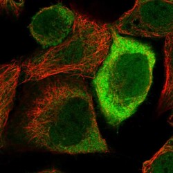
- Experimental details
- Immunofluorescent staining of human cell line A-431 shows localization to nucleoplasm, plasma membrane & cytosol.
- Sample type
- Human

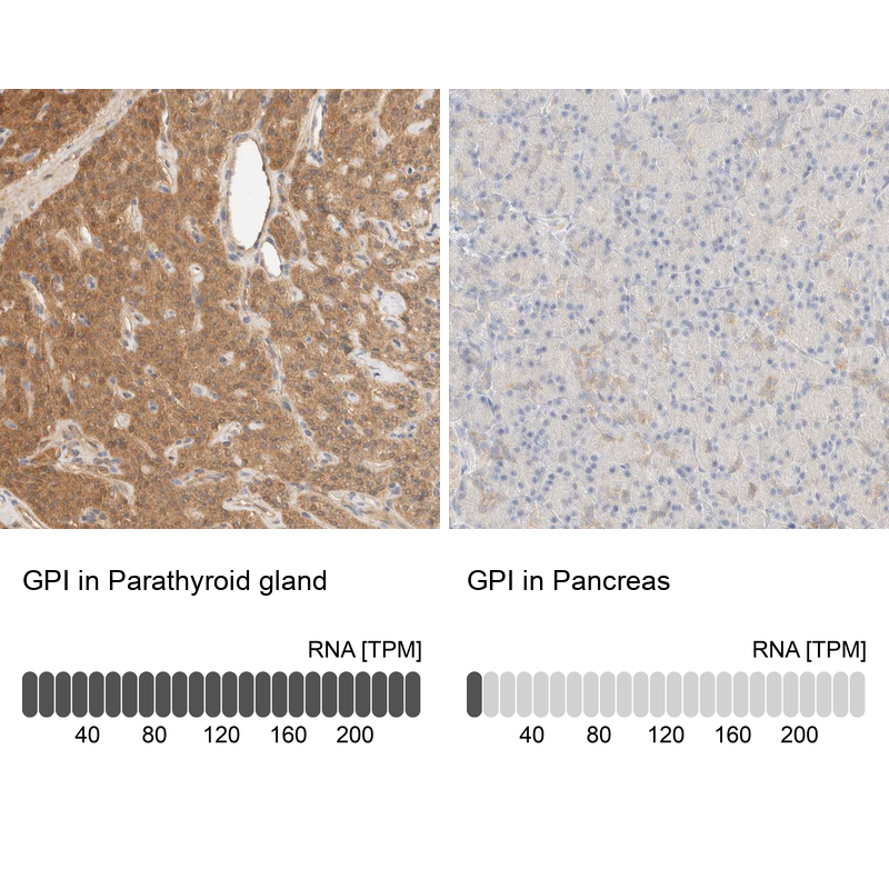
Supportive validation
- Submitted by
- Atlas Antibodies (provider)
- Enhanced method
- Orthogonal validation
- Main image

- Experimental details
- Immunohistochemistry analysis in human parathyroid gland and pancreas tissues using HPA024305 antibody. Corresponding GPI RNA-seq data are presented for the same tissues.
- Sample type
- Human
- Protocol
- Protocol