 Explore
 Validate
 Learn
Learn Western blot
 Immunohistochemistry
Antibody data
- Antibody Data
- Antigen structure
- References [3]
- Comments [0]
- Validations
- Immunohistochemistry [1]
Submit
Validation data
Reference
Comment
Report error
- Product number
- HPA022268 - Provider product page

- Provider
- Atlas Antibodies
- Proper citation
- Atlas Antibodies Cat#HPA022268, RRID:AB_1845225
- Product name
- Anti-LPCAT1
- Antibody type
- Polyclonal
- Description
- Polyclonal Antibody against Human LPCAT1, Gene description: lysophosphatidylcholine acyltransferase 1, Alternative Gene Names: AYTL2, FLJ12443, Validated applications: WB, IHC, Uniprot ID: Q8NF37, Storage: Store at +4°C for short term storage. Long time storage is recommended at -20°C.
- Reactivity
- Human
- Host
- Rabbit
- Conjugate
- Unconjugated
- Isotype
- IgG
- Vial size
- 100 µl
- Concentration
- 1.1 mg/ml
- Storage
- Store at +4°C for short term storage. Long time storage is recommended at -20°C.
- Handling
- The antibody solution should be gently mixed before use.
Submitted references Effects of Subretinal Gene Transfer at Different Time Points in a Mouse Model of Retinal Degeneration
AAV-Mediated Lysophosphatidylcholine Acyltransferase 1 (Lpcat1) Gene Replacement Therapy Rescues Retinal Degeneration inrd11Mice
Loss of lysophosphatidylcholine acyltransferase 1 leads to photoreceptor degeneration in rd11 mice
Swaroop A, Dai X, Zhang H, Han J, He Y, Zhang Y, Qi Y, Pang J
PLOS ONE 2016;11(5):e0156542
PLOS ONE 2016;11(5):e0156542
AAV-Mediated Lysophosphatidylcholine Acyltransferase 1 (Lpcat1) Gene Replacement Therapy Rescues Retinal Degeneration inrd11Mice
Dai X, Han J, Qi Y, Zhang H, Xiang L, Lv J, Li J, Deng W, Chang B, Hauswirth W, Pang J
Investigative Opthalmology & Visual Science 2014;55(3):1724
Investigative Opthalmology & Visual Science 2014;55(3):1724
Loss of lysophosphatidylcholine acyltransferase 1 leads to photoreceptor degeneration in rd11 mice
Friedman J, Chang B, Krauth D, Lopez I, Waseem N, Hurd R, Feathers K, Branham K, Shaw M, Thomas G, Brooks M, Liu C, Bakeri H, Campos M, Maubaret C, Webster A, Rodriguez I, Thompson D, Bhattacharya S, Koenekoop R, Heckenlively J, Swaroop A
Proceedings of the National Academy of Sciences 2010;107(35):15523-15528
Proceedings of the National Academy of Sciences 2010;107(35):15523-15528
No comments: Submit comment
Supportive validation
- Submitted by
- Atlas Antibodies (provider)
- Enhanced method
- Orthogonal validation
- Main image
- Experimental details
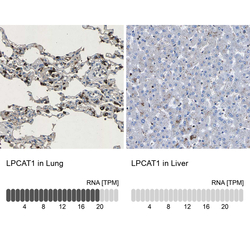
- Immunohistochemistry analysis in human lung and liver tissues using HPA022268 antibody. Corresponding LPCAT1 RNA-seq data are presented for the same tissues.
- Sample type
- Human
- Protocol
- Protocol
