 Explore
 Validate
 Learn
Learn Western blot
 ELISA
ELISAAntibody data
- Antibody Data
- Antigen structure
- References [10]
- Comments [0]
- Validations
- Western blot [1]
- Immunocytochemistry [1]
Submit
Validation data
Reference
Comment
Report error
- Product number
- 66044-1-Ig - Provider product page

- Provider
- Proteintech Group
- Proper citation
- Proteintech Cat#66044-1-Ig, RRID:AB_11045658
- Product name
- LPCAT1 antibody
- Antibody type
- Monoclonal
- Description
- KD/KO validated LPCAT1 antibody (Cat. #66044-1-Ig) is a mouse monoclonal antibody that shows reactivity with human, mouse, rat and has been validated for the following applications: IF, IHC, WB, ELISA.
- Reactivity
- Human, Mouse, Rat
- Host
- Mouse
- Conjugate
- Unconjugated
- Isotype
- IgG
- Antibody clone number
- 8B6E9
- Vial size
- 20ul, 150ul
Submitted references LPCAT1 as a prognostic biomarker and risk indicator for hepatocellular carcinoma: insights into genes related to lipid metabolism.
Chaihu Guizhi Ganjiang Decoction attenuates nonalcoholic steatohepatitis by enhancing intestinal barrier integrity and ameliorating PPARα mediated lipotoxicity.
LPCAT1 Facilitates Keratinocyte Hyperproliferation and Skin Inflammation in Psoriasis by Regulating GLUT3.
Stearoyl-CoA desaturase 1 deficiency exacerbates palmitate-induced lipotoxicity by the formation of small lipid droplets in pancreatic β-cells.
No evidence for carcinogenicity of titanium dioxide nanoparticles in 26-week inhalation study in rasH2 mouse model.
Lipid reprogramming induced by the TFEB-ERRα axis enhanced membrane fluidity to promote EC progression.
Gut Microbiota Dysbiosis Accelerates Prostate Cancer Progression Through Increased LPCAT1 Expression and Enhanced DNA Repair Pathways.
LPCAT1 reprogramming cholesterol metabolism promotes the progression of esophageal squamous cell carcinoma.
A miR-205-LPCAT1 axis contributes to proliferation and progression in multiple cancers.
Oncogene Amplification in Growth Factor Signaling Pathways Renders Cancers Dependent on Membrane Lipid Remodeling.
Zhai H, Wang J, He Y, Hong S, Huang K, Hu S, Xu J, Hao S, Zhang G, Shi X
Frontiers in oncology 2026;16:1707270
Frontiers in oncology 2026;16:1707270
Chaihu Guizhi Ganjiang Decoction attenuates nonalcoholic steatohepatitis by enhancing intestinal barrier integrity and ameliorating PPARα mediated lipotoxicity.
Wu H, Lou T, Pan M, Wei Z, Yang X, Liu L, Feng M, Shi L, Qu B, Cong S, Chen K, Yang H, Liu J, Li Y, Jia Z, Xiao H
Journal of ethnopharmacology 2024 May 23;326:117841
Journal of ethnopharmacology 2024 May 23;326:117841
LPCAT1 Facilitates Keratinocyte Hyperproliferation and Skin Inflammation in Psoriasis by Regulating GLUT3.
Huang Y, Wang Y, Zhen Y, Liu W, Wang Y, Wang R, Wang N, Huang S, Yan J, Sun Q
The Journal of investigative dermatology 2024 Jul;144(7):1479-1490.e14
The Journal of investigative dermatology 2024 Jul;144(7):1479-1490.e14
Stearoyl-CoA desaturase 1 deficiency exacerbates palmitate-induced lipotoxicity by the formation of small lipid droplets in pancreatic β-cells.
Janikiewicz J, Dobosz AM, Majzner K, Bernas T, Dobrzyn A
Biochimica et biophysica acta. Molecular basis of disease 2023 Aug;1869(6):166711
Biochimica et biophysica acta. Molecular basis of disease 2023 Aug;1869(6):166711
No evidence for carcinogenicity of titanium dioxide nanoparticles in 26-week inhalation study in rasH2 mouse model.
Yamano S, Takeda T, Goto Y, Hirai S, Furukawa Y, Kikuchi Y, Kasai T, Misumi K, Suzuki M, Takanobu K, Senoh H, Saito M, Kondo H, Umeda Y
Scientific reports 2022 Sep 2;12(1):14969
Scientific reports 2022 Sep 2;12(1):14969
Lipid reprogramming induced by the TFEB-ERRα axis enhanced membrane fluidity to promote EC progression.
Mao X, Lei H, Yi T, Su P, Tang S, Tong Y, Dong B, Ruan G, Mustea A, Sehouli J, Sun P
Journal of experimental & clinical cancer research : CR 2022 Jan 19;41(1):28
Journal of experimental & clinical cancer research : CR 2022 Jan 19;41(1):28
Gut Microbiota Dysbiosis Accelerates Prostate Cancer Progression Through Increased LPCAT1 Expression and Enhanced DNA Repair Pathways.
Liu Y, Yang C, Zhang Z, Jiang H
Frontiers in oncology 2021;11:679712
Frontiers in oncology 2021;11:679712
LPCAT1 reprogramming cholesterol metabolism promotes the progression of esophageal squamous cell carcinoma.
Tao M, Luo J, Gu T, Yu X, Song Z, Jun Y, Gu H, Han K, Huang X, Yu W, Sun S, Zhang Z, Liu L, Chen X, Zhang L, Luo C, Wang Q
Cell death & disease 2021 Sep 13;12(9):845
Cell death & disease 2021 Sep 13;12(9):845
A miR-205-LPCAT1 axis contributes to proliferation and progression in multiple cancers.
Liu F, Wu Y, Liu J, Ni RJ, Yang AG, Bian K, Zhang R
Biochemical and biophysical research communications 2020 Jun 25;527(2):474-480
Biochemical and biophysical research communications 2020 Jun 25;527(2):474-480
Oncogene Amplification in Growth Factor Signaling Pathways Renders Cancers Dependent on Membrane Lipid Remodeling.
Bi J, Ichu TA, Zanca C, Yang H, Zhang W, Gu Y, Chowdhry S, Reed A, Ikegami S, Turner KM, Zhang W, Villa GR, Wu S, Quehenberger O, Yong WH, Kornblum HI, Rich JN, Cloughesy TF, Cavenee WK, Furnari FB, Cravatt BF, Mischel PS
Cell metabolism 2019 Sep 3;30(3):525-538.e8
Cell metabolism 2019 Sep 3;30(3):525-538.e8
No comments: Submit comment
Supportive validation
- Submitted by
- Proteintech Group (provider)
- Main image
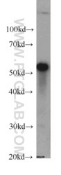
- Experimental details
- MCF7 cells were subjected to SDS PAGE followed by western blot with 66044-1-Ig(LPCAT1 antibody) at dilution of 1:500
- Sample type
- cell line

Supportive validation
- Submitted by
- Proteintech Group (provider)
- Main image

- Experimental details
- Immunofluorescent analysis of Hela cells, using LPCAT1 antibody 66044-1-lg at 1:50 dilution and Rhodamine-labeled goat anti-mouse IgG (red).
- Sample type
- cell line
